 Movie
Movie Controller

[English] 日本語
 Yorodumi
- PDB-2pgn: The crystal structure of FAD and ThDP-dependent Cyclohexane-1,2-d... -
+ Open data

- Basic information
| Entry | Database: PDB / ID: 2pgn | ||||||
|---|---|---|---|---|---|---|---|
| Title | The crystal structure of FAD and ThDP-dependent Cyclohexane-1,2-dione Hydrolase in Complex with Cyclohexane-1,2-dione | ||||||
 Components Components | Cyclohexane-1,2-dione Hydrolase (Cdh) | ||||||
 Keywords Keywords | HYDROLASE / Three alpha/beta domains | ||||||
| Function / homology | Thiamin diphosphate (ThDP)-binding fold, Pyr/PP domains / TPP-binding domain / Rossmann fold / 3-Layer(aba) Sandwich / Alpha Beta / CYCLOHEXANE-1,2-DIONE / FLAVIN-ADENINE DINUCLEOTIDE / THIAMINE DIPHOSPHATE Function and homology information Function and homology information | ||||||
| Biological species |  Azoarcus sp. (bacteria) Azoarcus sp. (bacteria) | ||||||
| Method |  X-RAY DIFFRACTION / SYNCHROTRON / known crystal structure / Resolution: 1.2 Å X-RAY DIFFRACTION / SYNCHROTRON / known crystal structure / Resolution: 1.2 Å | ||||||
 Authors Authors | Fraas, S. / Warkentin, E. / Ermler, U. | ||||||
 Citation Citation | Journal: To be Published Title: The crystal structure of FAD and ThDP-dependent Cyclohexane-1,2-dione Hydrolase in Complex with Cyclohexane-1,2-dione Authors: Fraas, S. / Steinbach, A.K. / Warkentin, E. / Kroneck, P.M.H. / Ermler, U. | ||||||
| History |
| ||||||
| Remark 999 | sequence The sequence is not available in UniProt database at the time of processing. |
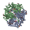
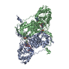
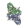
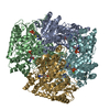
- Structure visualization
| Structure viewer | Molecule: MolmilJmol/JSmol |
|---|
- Downloads & links
-Download
| PDBx/mmCIF format | 2pgn.cif.gz | 480.9 KB | Display | PDBx/mmCIF format |
|---|---|---|---|---|
| PDB format | pdb2pgn.ent.gz | 388.8 KB | Display | PDB format |
| PDBx/mmJSON format | 2pgn.json.gz | Tree view | PDBx/mmJSON format | |
| Others |  Other downloads Other downloads |
-Validation report
| Arichive directory | https://data.pdbj.org/pub/pdb/validation_reports/pg/2pgnftp://data.pdbj.org/pub/pdb/validation_reports/pg/2pgn | HTTPS FTP |
|---|
-Related structure data
| Related structure data | |
|---|---|
| Similar structure data |


-Links
 PDBj
PDBj

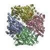
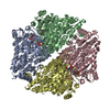
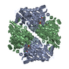
- Assembly
| Deposited unit | 
| |||||||||
|---|---|---|---|---|---|---|---|---|---|---|
| 1 | 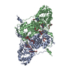
| |||||||||
| Unit cell |
| |||||||||
| Components on special symmetry positions |
|
-Components
-Protein , 1 types, 2 molecules AB
| #1: Protein | Mass: 63362.930 Da / Num. of mol.: 2 / Source method: isolated from a natural source / Source: (natural) Azoarcus sp. (bacteria) / Strain: 22Lin |
|---|
-Non-polymers , 6 types, 1285 molecules 


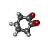

| #2: Chemical |  Mass: 24.305 Da / Num. of mol.: 2 / Source method: obtained synthetically / Formula: Mg Mass: 24.305 Da / Num. of mol.: 2 / Source method: obtained synthetically / Formula: Mg#3: Chemical | ChemComp-P6G /  Mass: 282.331 Da / Num. of mol.: 9 / Source method: obtained synthetically / Formula: C12H26O7 / Comment: precipitant*YM Mass: 282.331 Da / Num. of mol.: 9 / Source method: obtained synthetically / Formula: C12H26O7 / Comment: precipitant*YM#4: Chemical |  Mass: 785.550 Da / Num. of mol.: 2 / Source method: obtained synthetically / Formula: C27H33N9O15P2 / Comment: FAD*YM Mass: 785.550 Da / Num. of mol.: 2 / Source method: obtained synthetically / Formula: C27H33N9O15P2 / Comment: FAD*YM#5: Chemical |  Mass: 425.314 Da / Num. of mol.: 2 / Source method: obtained synthetically / Formula: C12H19N4O7P2S Mass: 425.314 Da / Num. of mol.: 2 / Source method: obtained synthetically / Formula: C12H19N4O7P2S#6: Chemical |  Mass: 112.127 Da / Num. of mol.: 2 / Source method: obtained synthetically / Formula: C6H8O2 Mass: 112.127 Da / Num. of mol.: 2 / Source method: obtained synthetically / Formula: C6H8O2#7: Water | ChemComp-HOH / | Mass: 18.015 Da / Num. of mol.: 1268 / Source method: isolated from a natural source / Formula: H2O |
|---|
-Experimental details
-Experiment
| Experiment | Method: X-RAY DIFFRACTION / Number of used crystals: 1 |
|---|
- Sample preparation
| Crystal | Density Matthews: 2.18 Å3/Da / Density % sol: 43.48 % |
|---|---|
| Crystal grow | Temperature: 298 K / Method: vapor diffusion, sitting drop / pH: 7 Details: 23% PEG 400, 0.01M sodium acetate, 3mM HEPES, 0.5mM ThDP, 0.5mM NAD, 0.1M NaCl. Crystal soaked in 47% PEG 400, 0.02M sodium acetate, 5mM 1,2 cyclohexanedione, pH 7.0, VAPOR DIFFUSION, ...Details: 23% PEG 400, 0.01M sodium acetate, 3mM HEPES, 0.5mM ThDP, 0.5mM NAD, 0.1M NaCl. Crystal soaked in 47% PEG 400, 0.02M sodium acetate, 5mM 1,2 cyclohexanedione, pH 7.0, VAPOR DIFFUSION, SITTING DROP, temperature 298K |
-Data collection
| Diffraction | Mean temperature: 100 K |
|---|---|
| Diffraction source | Source: SYNCHROTRON / Site: SLS  / Beamline: X10SA / Wavelength: 1 Å / Beamline: X10SA / Wavelength: 1 Å |
| Detector | Type: MARMOSAIC 225 mm CCD / Detector: CCD / Date: Mar 3, 2005 |
| Radiation | Monochromator: Si(111) / Protocol: SINGLE WAVELENGTH / Monochromatic (M) / Laue (L): M / Scattering type: x-ray |
| Radiation wavelength | Wavelength: 1 Å / Relative weight: 1 |
| Reflection | Resolution: 1.1→40 Å / Num. all: 388867 / Num. obs: 379045 / % possible obs: 84.8 % / Observed criterion σ(F): 0 / Observed criterion σ(I): 0 / Redundancy: 6.1 % / Biso Wilson estimate: 12.26 Å2 / Rmerge(I) obs: 0.068 / Net I/σ(I): 13.58 |
| Reflection shell | Resolution: 1.1→1.2 Å / Redundancy: 3.6 % / Rmerge(I) obs: 0.487 / Mean I/σ(I) obs: 2.7 / Num. measured obs: 187829 / Num. unique all: 50802 / % possible all: 49.9 |
- Processing
| Software |
| ||||||||||||||||||||||||||||||||||||||||||||||||||||||||||||||||||||||||||||||||||||||||||||||||||||||||||||||||||||||||||||||||||||||||||||
|---|---|---|---|---|---|---|---|---|---|---|---|---|---|---|---|---|---|---|---|---|---|---|---|---|---|---|---|---|---|---|---|---|---|---|---|---|---|---|---|---|---|---|---|---|---|---|---|---|---|---|---|---|---|---|---|---|---|---|---|---|---|---|---|---|---|---|---|---|---|---|---|---|---|---|---|---|---|---|---|---|---|---|---|---|---|---|---|---|---|---|---|---|---|---|---|---|---|---|---|---|---|---|---|---|---|---|---|---|---|---|---|---|---|---|---|---|---|---|---|---|---|---|---|---|---|---|---|---|---|---|---|---|---|---|---|---|---|---|---|---|---|
| Refinement | Method to determine structure: known crystal structure / Resolution: 1.2→10 Å / Cor.coef. Fo:Fc: 0.978 / Cor.coef. Fo:Fc free: 0.97 / SU B: 1.52 / SU ML: 0.03 / Cross valid method: THROUGHOUT / σ(F): 0 / ESU R: 0.04 / ESU R Free: 0.04 / Stereochemistry target values: MAXIMUM LIKELIHOOD / Details: HYDROGENS HAVE BEEN ADDED IN THE RIDING POSITIONS
| ||||||||||||||||||||||||||||||||||||||||||||||||||||||||||||||||||||||||||||||||||||||||||||||||||||||||||||||||||||||||||||||||||||||||||||
| Solvent computation | Ion probe radii: 0.8 Å / Shrinkage radii: 0.8 Å / VDW probe radii: 1.2 Å / Solvent model: MASK | ||||||||||||||||||||||||||||||||||||||||||||||||||||||||||||||||||||||||||||||||||||||||||||||||||||||||||||||||||||||||||||||||||||||||||||
| Displacement parameters | Biso mean: 12.78 Å2
| ||||||||||||||||||||||||||||||||||||||||||||||||||||||||||||||||||||||||||||||||||||||||||||||||||||||||||||||||||||||||||||||||||||||||||||
| Refinement step | Cycle: LAST / Resolution: 1.2→10 Å
| ||||||||||||||||||||||||||||||||||||||||||||||||||||||||||||||||||||||||||||||||||||||||||||||||||||||||||||||||||||||||||||||||||||||||||||
| Refine LS restraints |
| ||||||||||||||||||||||||||||||||||||||||||||||||||||||||||||||||||||||||||||||||||||||||||||||||||||||||||||||||||||||||||||||||||||||||||||
| LS refinement shell | Resolution: 1.2→1.231 Å / Total num. of bins used: 20
|
