 Movie
Movie Controller


+ Open data

- Basic information
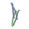
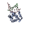
| Entry | Database: PDB / ID: 2fyz | ||||||
|---|---|---|---|---|---|---|---|
| Title | Structural of Mumps virus fusion protein core | ||||||
 Components Components | (Fusion glycoprotein F0) x 2 | ||||||
 Keywords Keywords | PROTEIN BINDING / Mumps virus fusion protein core | ||||||
| Function / homology |  Function and homology information Function and homology informationmembrane => GO:0016020 / fusion of virus membrane with host plasma membrane / viral envelope / host cell plasma membrane / virion membrane Similarity search - Function | ||||||
| Biological species |   Mumps virus Mumps virus | ||||||
| Method |  X-RAY DIFFRACTION / MOLECULAR REPLACEMENT / Resolution: 2.2 Å X-RAY DIFFRACTION / MOLECULAR REPLACEMENT / Resolution: 2.2 Å | ||||||
 Authors Authors | Lou, Z. / Xu, Y. / Liu, Y. / Rao, Z. | ||||||
 Citation Citation | Journal: Biochem.Biophys.Res.Commun. / Year: 2006 Title: Structural characterization of mumps virus fusion protein core Authors: Liu, Y. / Xu, Y. / Lou, Z. / Zhu, J. / Hu, X. / Gao, G.F. / Qiu, B. / Rao, Z. / Tien, P. | ||||||
| History |
|
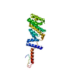
- Structure visualization
| Structure viewer | Molecule: MolmilJmol/JSmol |
|---|
- Downloads & links
-Download
| PDBx/mmCIF format | 2fyz.cif.gz | 70 KB | Display | PDBx/mmCIF format |
|---|---|---|---|---|
| PDB format | pdb2fyz.ent.gz | 52.5 KB | Display | PDB format |
| PDBx/mmJSON format | 2fyz.json.gz | Tree view | PDBx/mmJSON format | |
| Others |  Other downloads Other downloads |
-Validation report
| Summary document | 2fyz_validation.pdf.gz | 468.4 KB | Display | wwPDB validaton report |
|---|---|---|---|---|
| Full document | 2fyz_full_validation.pdf.gz | 478 KB | Display | |
| Data in XML | 2fyz_validation.xml.gz | 16.3 KB | Display | |
| Data in CIF | 2fyz_validation.cif.gz | 22.8 KB | Display | |
| Arichive directory | https://data.pdbj.org/pub/pdb/validation_reports/fy/2fyzftp://data.pdbj.org/pub/pdb/validation_reports/fy/2fyz | HTTPS FTP |
-Related structure data
| Related structure data |  1svfS S: Starting model for refinement |
|---|---|
| Similar structure data |


-Links
 PDBj
PDBj

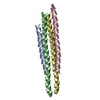
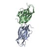
- Assembly
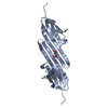
| Deposited unit | 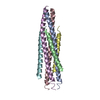
| ||||||||
|---|---|---|---|---|---|---|---|---|---|
| 1 |
| ||||||||
| Unit cell |
| ||||||||
| Details | There are 3 molecules in one ASU. They form the biological unit. |
-Components
| #1: Protein | Mass: 6694.612 Da / Num. of mol.: 3 / Fragment: residues 119-181 Source method: isolated from a genetically manipulated source Source: (gene. exp.) Mumps virus / Genus: Rubulavirus / Plasmid: pGEX-6p-1 / Species (production host): Escherichia coli / Production host:  #2: Protein/peptide | Mass: 5080.690 Da / Num. of mol.: 3 / Fragment: residues 438-485 / Mutation: A463T, N478I Source method: isolated from a genetically manipulated source Source: (gene. exp.) Mumps virus / Genus: Rubulavirus / Plasmid: pGEX-6p-1 / Species (production host): Escherichia coli / Production host: #3: Water | ChemComp-HOH / |  Mass: 18.015 Da / Num. of mol.: 263 / Source method: isolated from a natural source / Formula: H2O Mass: 18.015 Da / Num. of mol.: 263 / Source method: isolated from a natural source / Formula: H2O |
|---|
-Experimental details
-Experiment
| Experiment | Method: X-RAY DIFFRACTION / Number of used crystals: 1 |
|---|
- Sample preparation
| Crystal | Density Matthews: 2.75 Å3/Da / Density % sol: 55.35 % |
|---|---|
| Crystal grow | Temperature: 291 K / Method: vapor diffusion, hanging drop / pH: 8 Details: 15% PEG 800, 0.5M Lithium Sulfate, pH 8.0, VAPOR DIFFUSION, HANGING DROP, temperature 291K |
-Data collection
| Diffraction | Mean temperature: 100 K |
|---|---|
| Diffraction source | Source: ROTATING ANODE / Type: RIGAKU RU200 / Wavelength: 1.5418 Å |
| Detector | Type: MAR scanner 345 mm plate / Detector: IMAGE PLATE |
| Radiation | Monochromator: osmic mirror / Protocol: SINGLE WAVELENGTH / Monochromatic (M) / Laue (L): M / Scattering type: x-ray |
| Radiation wavelength | Wavelength: 1.5418 Å / Relative weight: 1 |
| Reflection | Resolution: 2.2→50 Å / Num. all: 19670 / Num. obs: 17183 |
| Reflection shell | Resolution: 2.2→2.3 Å |
- Processing
| Software |
| ||||||||||||||||||||
|---|---|---|---|---|---|---|---|---|---|---|---|---|---|---|---|---|---|---|---|---|---|
| Refinement | Method to determine structure: MOLECULAR REPLACEMENT Starting model: 1SVF Resolution: 2.2→50 Å / σ(F): 0 / Stereochemistry target values: Engh & Huber
| ||||||||||||||||||||
| Refinement step | Cycle: LAST / Resolution: 2.2→50 Å
| ||||||||||||||||||||
| Refine LS restraints |
|
