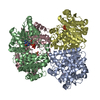
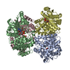
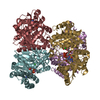
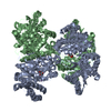
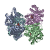
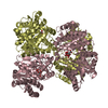
Entry Database : PDB / ID : 2bo4Title Dissection of mannosylglycerate synthase: an archetypal mannosyltransferase MANNOSYLGLYCERATE SYNTHASE Keywords / / / / Function / homology Function Domain/homology Component
/ / / / / / / / / / / / / Biological species RHODOTHERMUS MARINUS (bacteria)Method / / / Resolution : 1.95 Å Authors Flint, J. / Taylor, E. / Yang, M. / Bolam, D.N. / Tailford, L.E. / Martinez-Fleites, C. / Dodson, E.J. / Davis, B.G. / Gilbert, H.J. / Davies, G.J. Journal : Nat.Struct.Mol.Biol. / Year : 2005Title : Structural Dissection and High-Throughput Screening of Mannosylglyceerate SynthaseAuthors : Flint, J. / Taylor, E. / Yang, M. / Bolam, D.N. / Tailford, L.E. / Martinez-Fleites, C. / Dodson, E.J. / Davis, B.G. / Gilbert, H.J. / Davies, G.J. History Deposition Apr 7, 2005 Deposition site / Processing site Revision 1.0 Jun 6, 2005 Provider / Type Revision 1.1 Jul 13, 2011 Group / Refinement description / Version format complianceRevision 1.2 May 8, 2024 Group Data collection / Database references ... Data collection / Database references / Derived calculations / Other Category chem_comp_atom / chem_comp_bond ... chem_comp_atom / chem_comp_bond / database_2 / pdbx_database_status / struct_site Item _database_2.pdbx_DOI / _database_2.pdbx_database_accession ... _database_2.pdbx_DOI / _database_2.pdbx_database_accession / _pdbx_database_status.status_code_sf / _struct_site.pdbx_auth_asym_id / _struct_site.pdbx_auth_comp_id / _struct_site.pdbx_auth_seq_id
Show all Show less
 Movie
Movie Controller

 Yorodumi
 Open data
 Basic information
 Components
 Keywords
 Function and homology information
 RHODOTHERMUS MARINUS (bacteria)
 X-RAY DIFFRACTION /
  Authors
 Citation
 Structure visualization
 Downloads & links
 Other downloads


 PDBj
PDBj


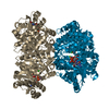
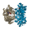
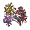
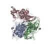
 Assembly
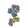

 Mass: 189.100 Da / Num. of mol.: 6 / Source method: obtained synthetically / Formula: C6H5O7
 Mass: 18.015 Da / Num. of mol.: 1767 / Source method: isolated from a natural source / Formula: H2O
 Sample preparation
 / Beamline: ID14-4 / Wavelength: 0.9783
  Processing