 Movie
Movie Controller


+ Open data

- Basic information
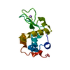
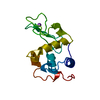
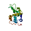
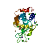
| Entry | Database: PDB / ID: 208l | ||||||
|---|---|---|---|---|---|---|---|
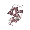
| Title | MUTANT HUMAN LYSOZYME C77A | ||||||
 Components Components | LYSOZYME | ||||||
 Keywords Keywords | COMPLEX (HYDROLASE (O-GLYCOSYL)/CYS) / COMPLEX (HYDROLASE (O-GLYCOSYL)-CYS) / MUTANT HUMAN LYSOZYME / HYDROLASE / COMPLEX (HYDROLASE (O-GLYCOSYL)-CYS) complex | ||||||
| Function / homology |  Function and homology informationcytolysis / antimicrobial humoral response / retina homeostasis / Antimicrobial peptides / metabolic process / specific granule lumen / azurophil granule lumen / tertiary granule lumen / lysozyme / lysozyme activity ...cytolysis / antimicrobial humoral response / retina homeostasis / Antimicrobial peptides / metabolic process / specific granule lumen / azurophil granule lumen / tertiary granule lumen / lysozyme / lysozyme activity / killing of cells of another organism / defense response to Gram-negative bacterium / defense response to Gram-positive bacterium / defense response to bacterium / inflammatory response / Amyloid fiber formation / Neutrophil degranulation / extracellular space / extracellular exosome / extracellular region / identical protein binding Function and homology informationcytolysis / antimicrobial humoral response / retina homeostasis / Antimicrobial peptides / metabolic process / specific granule lumen / azurophil granule lumen / tertiary granule lumen / lysozyme / lysozyme activity ...cytolysis / antimicrobial humoral response / retina homeostasis / Antimicrobial peptides / metabolic process / specific granule lumen / azurophil granule lumen / tertiary granule lumen / lysozyme / lysozyme activity / killing of cells of another organism / defense response to Gram-negative bacterium / defense response to Gram-positive bacterium / defense response to bacterium / inflammatory response / Amyloid fiber formation / Neutrophil degranulation / extracellular space / extracellular exosome / extracellular region / identical protein bindingSimilarity search - Function | ||||||
| Biological species |  Homo sapiens (human) Homo sapiens (human) | ||||||
| Method | X-RAY DIFFRACTION / MOLECULAR REPLACEMENT / Resolution: 2.2 Å | ||||||
 Authors Authors | Matsushima, M. / Song, H. | ||||||
 Citation Citation | Journal: J.Biochem.(Tokyo) / Year: 1996 Title: A role of PDI in the reductive cleavage of mixed disulfides. Authors: Nakamura, S. / Matsushima, M. / Song, H. / Kikuchi, M. #1: Journal: Acta Crystallogr.,Sect.D / Year: 1995Title: Structure of a Glutathionylated Human Lysozyme: A Folding Intermediate Mimic in the Formation of a Disulfide Bond Authors: Inaka, K. / Miki, K. / Kikuchi, M. / Taniyama, Y. / Matsushima, M. #2: Journal: FEBS Lett. / Year: 1993Title: Pdi and Glutathione-Mediated Reduction of the Glutathionylated Variant of Human Lysozyme Authors: Hayano, T. / Inaka, K. / Otsu, M. / Taniyama, Y. / Miki, K. / Matsushima, M. / Kikuchi, M. | ||||||
| History |
|
- Structure visualization
| Structure viewer | Molecule: MolmilJmol/JSmol |
|---|
- Downloads & links
-Download
| PDBx/mmCIF format | 208l.cif.gz | 37.6 KB | Display | PDBx/mmCIF format |
|---|---|---|---|---|
| PDB format | pdb208l.ent.gz | 25.6 KB | Display | PDB format |
| PDBx/mmJSON format | 208l.json.gz | Tree view | PDBx/mmJSON format | |
| Others |  Other downloads Other downloads |
-Validation report
| Arichive directory | https://data.pdbj.org/pub/pdb/validation_reports/08/208lftp://data.pdbj.org/pub/pdb/validation_reports/08/208l | HTTPS FTP |
|---|
-Related structure data


-Links
 PDBj
PDBj


- Assembly
| Deposited unit | 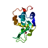
| ||||||||
|---|---|---|---|---|---|---|---|---|---|
| 1 |
| ||||||||
| Unit cell |
|
-Components
| #1: Protein | / HL_95CYS Mass: 14688.627 Da / Num. of mol.: 1 / Mutation: C77A Source method: isolated from a genetically manipulated source Source: (gene. exp.) Homo sapiens (human) / Production host:  Escherichia coli (E. coli) / References: UniProt: P00695, UniProt: P61626*PLUS, lysozyme Escherichia coli (E. coli) / References: UniProt: P00695, UniProt: P61626*PLUS, lysozyme |
|---|---|
| #2: Chemical | ChemComp-CYS / Cysteine  Type: L-peptide linking / Mass: 121.158 Da / Num. of mol.: 1 / Source method: obtained synthetically / Formula: C3H7NO2S Type: L-peptide linking / Mass: 121.158 Da / Num. of mol.: 1 / Source method: obtained synthetically / Formula: C3H7NO2SDetails: C77 CHANGED TO ALA AND A CYSTEINE WAS CHEMICALLY BOUND ON THE SH GROUP OF CYS95 THROUGH A DISULFIDE BOND |
| #3: Water | ChemComp-HOH / Water Mass: 18.015 Da / Num. of mol.: 40 / Source method: isolated from a natural source / Formula: H2O Mass: 18.015 Da / Num. of mol.: 40 / Source method: isolated from a natural source / Formula: H2O |
-Experimental details
-Experiment
| Experiment | Method: X-RAY DIFFRACTION / Number of used crystals: 1 |
|---|
- Sample preparation
| Crystal | Density Matthews: 1.96 Å3/Da / Density % sol: 37.27 % | ||||||||||||||||||||||||||||||||||||
|---|---|---|---|---|---|---|---|---|---|---|---|---|---|---|---|---|---|---|---|---|---|---|---|---|---|---|---|---|---|---|---|---|---|---|---|---|---|
| Crystal grow | pH: 6 / Details: pH 6.0 | ||||||||||||||||||||||||||||||||||||
| Crystal grow | *PLUS Temperature: 4 ℃ / pH: 4 / Method: vapor diffusion | ||||||||||||||||||||||||||||||||||||
| Components of the solutions | *PLUS
|
-Data collection
| Diffraction | Mean temperature: 280 K |
|---|---|
| Diffraction source | Source: ROTATING ANODE / Type: MACSCIENCE M18X / Wavelength: 1.5418 |
| Detector | Type: MACSCIENCE / Detector: IMAGE PLATE / Date: Feb 1, 1993 / Details: MIRROR-MIRROR |
| Radiation | Monochromatic (M) / Laue (L): M / Scattering type: x-ray |
| Radiation wavelength | Wavelength: 1.5418 Å / Relative weight: 1 |
| Reflection | Resolution: 1.8→100 Å / Observed criterion σ(I): 2 |
- Processing
| Software |
| ||||||||||||||||||||||||||||||||||||||||||||||||||||||||||||
|---|---|---|---|---|---|---|---|---|---|---|---|---|---|---|---|---|---|---|---|---|---|---|---|---|---|---|---|---|---|---|---|---|---|---|---|---|---|---|---|---|---|---|---|---|---|---|---|---|---|---|---|---|---|---|---|---|---|---|---|---|---|
| Refinement | Method to determine structure: MOLECULAR REPLACEMENT Starting model: NATIVE HUMAN LYSOZYME Resolution: 2.2→6 Å / σ(F): 2 /
| ||||||||||||||||||||||||||||||||||||||||||||||||||||||||||||
| Refinement step | Cycle: LAST / Resolution: 2.2→6 Å
| ||||||||||||||||||||||||||||||||||||||||||||||||||||||||||||
| Refine LS restraints |
| ||||||||||||||||||||||||||||||||||||||||||||||||||||||||||||
| Xplor file |
| ||||||||||||||||||||||||||||||||||||||||||||||||||||||||||||
| Software | *PLUS Name: X-PLOR / Classification: refinement | ||||||||||||||||||||||||||||||||||||||||||||||||||||||||||||
| Refinement | *PLUS | ||||||||||||||||||||||||||||||||||||||||||||||||||||||||||||
| Solvent computation | *PLUS | ||||||||||||||||||||||||||||||||||||||||||||||||||||||||||||
| Displacement parameters | *PLUS |
