 Movie
Movie Controller

[English] 日本語
 Yorodumi
- PDB-1yrg: THE CRYSTAL STRUCTURE OF RNA1P: A NEW FOLD FOR A GTPASE-ACTIVATIN... -
+ Open data

- Basic information
| Entry | Database: PDB / ID: 1yrg | ||||||
|---|---|---|---|---|---|---|---|
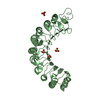
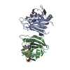
| Title | THE CRYSTAL STRUCTURE OF RNA1P: A NEW FOLD FOR A GTPASE-ACTIVATING PROTEIN | ||||||
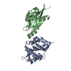
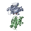
 Components Components | GTPASE-ACTIVATING PROTEIN RNA1_SCHPO | ||||||
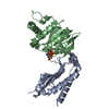
 Keywords Keywords | TRANSCRIPTION / GTPASE-ACTIVATING PROTEIN / GAP / RNA1P / RANGAP / LRR / LEUCINE-RICH REPEAT PROTEIN / TWINNING / HEMIHEDRAL TWINNING / MEROHEDRAL TWINNING / MEROHEDRY | ||||||
| Function / homology |  Function and homology information Function and homology informationSUMOylation of nuclear envelope proteins / Postmitotic nuclear pore complex (NPC) reformation / nuclear export / protein export from nucleus / nuclear periphery / GTPase activator activity / small GTPase binding / perinuclear region of cytoplasm / nucleus / cytoplasm / cytosol Similarity search - Function | ||||||
| Biological species |  | ||||||
| Method |  X-RAY DIFFRACTION / SYNCHROTRON / MIRAS / Resolution: 2.66 Å X-RAY DIFFRACTION / SYNCHROTRON / MIRAS / Resolution: 2.66 Å | ||||||
 Authors Authors | Hillig, R.C. / Renault, L. / Vetter, I.R. / Drell, T. / Wittinghofer, A. / Becker, J. | ||||||
 Citation Citation | Journal: Mol.Cell / Year: 1999 Title: The crystal structure of rna1p: a new fold for a GTPase-activating protein. Authors: Hillig, R.C. / Renault, L. / Vetter, I.R. / Drell 4th, T. / Wittinghofer, A. / Becker, J. #1: Journal: J.Biol.Chem. / Year: 1995 Title: RNA1 encodes a GTPase-activating protein specific for Gsp1p, the Ran/TC4 homologue of Saccharomyces cerevisiae. Authors: Becker, J. / Melchior, F. / Gerke, V. / Bischoff, F.R. / Ponstingl, H. / Wittinghofer, A. #2: Journal: Mol.Biol.Cell / Year: 1993 Title: A functional homologue of the RNA1 gene product in Schizosaccharomyces pombe: purification, biochemical characterization, and identification of a leucine-rich repeat motif. Authors: Melchior, F. / Weber, K. / Gerke, V. | ||||||
| History |
|
- Structure visualization
| Structure viewer | Molecule: MolmilJmol/JSmol |
|---|
- Downloads & links
-Download
| PDBx/mmCIF format | 1yrg.cif.gz | 142.2 KB | Display | PDBx/mmCIF format |
|---|---|---|---|---|
| PDB format | pdb1yrg.ent.gz | 112.9 KB | Display | PDB format |
| PDBx/mmJSON format | 1yrg.json.gz | Tree view | PDBx/mmJSON format | |
| Others |  Other downloads Other downloads |
-Validation report
| Arichive directory | https://data.pdbj.org/pub/pdb/validation_reports/yr/1yrgftp://data.pdbj.org/pub/pdb/validation_reports/yr/1yrg | HTTPS FTP |
|---|
-Related structure data
| Similar structure data |
|---|


-Links
 PDBj
PDBj- Assembly
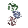
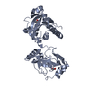
| Deposited unit | 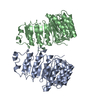
| ||||||||||
|---|---|---|---|---|---|---|---|---|---|---|---|
| 1 | 
| ||||||||||
| 2 | 
| ||||||||||
| Unit cell |
| ||||||||||
| Noncrystallographic symmetry (NCS) | NCS oper: (Code: given Matrix: (-0.99602, 0.08914, 0.00232), Vector: |
-Components
| #1: Protein | Mass: 43141.461 Da / Num. of mol.: 2 / Mutation: SER2ALA Source method: isolated from a genetically manipulated source Source: (gene. exp.) Description: SELENOMETHIONINE MUTANT EXPRESSED IN E.COLI B834 Plasmid: PET3D / Species (production host): Escherichia coli / Production host:  #2: Water | ChemComp-HOH / |  Mass: 18.015 Da / Num. of mol.: 21 / Source method: isolated from a natural source / Formula: H2O Mass: 18.015 Da / Num. of mol.: 21 / Source method: isolated from a natural source / Formula: H2OCompound details | THE ELEVEN LEUCINE-RICH REPEATS (LRR) OF S.POMBE RNA1P: LRR1(2-32), LRR2(33-60), LRR3(61-94), ...THE ELEVEN LEUCINE-RICH REPEATS (LRR) OF S.POMBE RNA1P: LRR1(2-32), LRR2(33-60), LRR3(61-94), LRR4(95-122), LRR5 (123-159), LRR6(160-187), LRR7(188-216), LRR8(217-244), LRR9(245-274),LRR10(275-303),LRR11(304-333). | Sequence details | MET1 CLEAVED OFF; MUTATION SER2ALA DUE TO EXPRESSION | |
|---|
-Experimental details
-Experiment
| Experiment | Method: X-RAY DIFFRACTION / Number of used crystals: 1 |
|---|
- Sample preparation
| Crystal | Density Matthews: 2.49 Å3/Da / Density % sol: 50.3 % Description: DATA SHOWED HEMIHEDRAL TWINNING. INTENSITIES WERE DE-TEWINNED FOR STRUCTURE DETERMINATION | ||||||||||||||||||||||||||||||||||||||||||||||||
|---|---|---|---|---|---|---|---|---|---|---|---|---|---|---|---|---|---|---|---|---|---|---|---|---|---|---|---|---|---|---|---|---|---|---|---|---|---|---|---|---|---|---|---|---|---|---|---|---|---|
| Crystal grow | Method: vapor diffusion, hanging drop / pH: 8.5 Details: HANGING DROPS MADE FROM 2-4 MICROLITER OF PROTEIN SOLUTION (25 MG/ML RNA1P IN 0.02 M TRIS-HCL, 0.002 M DTE, PH 7.5) AND THE SAME VOLUME OF RESERVOIR SOLUTION (21-24% PEG 2000 MME, 0.1 M TRIS- ...Details: HANGING DROPS MADE FROM 2-4 MICROLITER OF PROTEIN SOLUTION (25 MG/ML RNA1P IN 0.02 M TRIS-HCL, 0.002 M DTE, PH 7.5) AND THE SAME VOLUME OF RESERVOIR SOLUTION (21-24% PEG 2000 MME, 0.1 M TRIS-HCL, 0.2 M LI2SO4, 0.02 M MGCL2, PH 8.5), VAPOR DIFFUSION, HANGING DROP | ||||||||||||||||||||||||||||||||||||||||||||||||
| Crystal | *PLUS | ||||||||||||||||||||||||||||||||||||||||||||||||
| Crystal grow | *PLUS Temperature: 20 ℃ / pH: 7.5 | ||||||||||||||||||||||||||||||||||||||||||||||||
| Components of the solutions | *PLUS
|
-Data collection
| Diffraction | Mean temperature: 100 K |
|---|---|
| Diffraction source | Source: SYNCHROTRON / Site: ESRF  / Beamline: BM14 / Wavelength: 0.946 / Beamline: BM14 / Wavelength: 0.946 |
| Detector | Type: MARRESEARCH / Detector: IMAGE PLATE / Date: Jul 15, 1997 |
| Radiation | Protocol: SINGLE WAVELENGTH / Monochromatic (M) / Laue (L): M / Scattering type: x-ray |
| Radiation wavelength | Wavelength: 0.946 Å / Relative weight: 1 |
| Reflection | Resolution: 2.64→20 Å / Num. obs: 24708 / % possible obs: 97.8 % / Redundancy: 6.1 % / Biso Wilson estimate: 34 Å2 / Rsym value: 8.4 / Net I/σ(I): 18.2 |
| Reflection shell | Resolution: 2.64→2.69 Å / Mean I/σ(I) obs: 4.2 / Rsym value: 19.6 / % possible all: 58.9 |
| Reflection | *PLUS Rmerge(I) obs: 0.084 |
| Reflection shell | *PLUS % possible obs: 58.9 % / Rmerge(I) obs: 0.196 |
- Processing
| Software |
| ||||||||||||||||||||||||||||||||||||||||||||||||||||||||||||
|---|---|---|---|---|---|---|---|---|---|---|---|---|---|---|---|---|---|---|---|---|---|---|---|---|---|---|---|---|---|---|---|---|---|---|---|---|---|---|---|---|---|---|---|---|---|---|---|---|---|---|---|---|---|---|---|---|---|---|---|---|---|
| Refinement | Method to determine structure: MIRAS / Resolution: 2.66→20 Å / Rfactor Rfree error: 0.008 / Data cutoff high rms absF: 2684368.83 / Isotropic thermal model: RESTRAINED / Cross valid method: THROUGHOUT / σ(F): 0 Details: REFINEMENT TARGET: MAXIMUM LIKELIHOOD TARGET USING AMPLITUDES
| ||||||||||||||||||||||||||||||||||||||||||||||||||||||||||||
| Solvent computation | Solvent model: FLAT MODEL / Bsol: 19.03 Å2 / ksol: 0.34 e/Å3 | ||||||||||||||||||||||||||||||||||||||||||||||||||||||||||||
| Displacement parameters | Biso mean: 22.5 Å2
| ||||||||||||||||||||||||||||||||||||||||||||||||||||||||||||
| Refine analyze |
| ||||||||||||||||||||||||||||||||||||||||||||||||||||||||||||
| Refinement step | Cycle: LAST / Resolution: 2.66→20 Å
| ||||||||||||||||||||||||||||||||||||||||||||||||||||||||||||
| Refine LS restraints |
| ||||||||||||||||||||||||||||||||||||||||||||||||||||||||||||
| LS refinement shell | Resolution: 2.66→2.83 Å / Rfactor Rfree error: 0.03 / Total num. of bins used: 6
| ||||||||||||||||||||||||||||||||||||||||||||||||||||||||||||
| Xplor file |
| ||||||||||||||||||||||||||||||||||||||||||||||||||||||||||||
| Software | *PLUS Name: CNS / Version: 0.4 / Classification: refinement | ||||||||||||||||||||||||||||||||||||||||||||||||||||||||||||
| Refinement | *PLUS Lowest resolution: 20 Å / σ(F): 0 / % reflection Rfree: 5.1 % / Rfactor obs: 0.228 | ||||||||||||||||||||||||||||||||||||||||||||||||||||||||||||
| Solvent computation | *PLUS | ||||||||||||||||||||||||||||||||||||||||||||||||||||||||||||
| Displacement parameters | *PLUS Biso mean: 22.5 Å2 | ||||||||||||||||||||||||||||||||||||||||||||||||||||||||||||
| Refine LS restraints | *PLUS
| ||||||||||||||||||||||||||||||||||||||||||||||||||||||||||||
| LS refinement shell | *PLUS Rfactor Rfree: 0.373 / % reflection Rfree: 5 % / Rfactor Rwork: 0.299 |
