 Movie
Movie Controller


+ Open data

- Basic information
| Entry | Database: PDB / ID: 1sq5 | ||||||
|---|---|---|---|---|---|---|---|
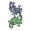
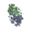
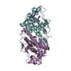
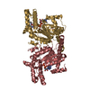
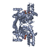
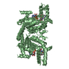
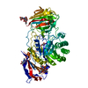
| Title | Crystal Structure of E. coli Pantothenate kinase | ||||||
 Components Components | Pantothenate kinase | ||||||
 Keywords Keywords | TRANSFERASE / P-loop | ||||||
| Function / homology |  Function and homology information Function and homology informationpantetheine kinase activity / pantothenate kinase / pantothenate kinase activity / coenzyme A biosynthetic process / protein homodimerization activity / ATP binding / cytoplasm Similarity search - Function | ||||||
| Biological species |  | ||||||
| Method |  X-RAY DIFFRACTION / SYNCHROTRON / MOLECULAR REPLACEMENT / Resolution: 2.2 Å X-RAY DIFFRACTION / SYNCHROTRON / MOLECULAR REPLACEMENT / Resolution: 2.2 Å | ||||||
 Authors Authors | Ivey, R.A. / Zhang, Y.-M. / Virga, K.G. / Hevener, K. / Lee, R.E. / Rock, C.O. / Jackowski, S. / Park, H.-W. | ||||||
 Citation Citation | Journal: J.Biol.Chem. / Year: 2004 Title: The structure of the pantothenate kinase.ADP.pantothenate ternary complex reveals the relationship between the binding sites for substrate, allosteric regulator, and antimetabolites. Authors: Ivey, R.A. / Zhang, Y.-M. / Virga, K.G. / Hevener, K. / Lee, R.E. / Rock, C.O. / Jackowski, S. / Park, H.-W. | ||||||
| History |
|
- Structure visualization
| Structure viewer | Molecule: MolmilJmol/JSmol |
|---|
- Downloads & links
-Download
| PDBx/mmCIF format | 1sq5.cif.gz | 276.6 KB | Display | PDBx/mmCIF format |
|---|---|---|---|---|
| PDB format | pdb1sq5.ent.gz | 217.7 KB | Display | PDB format |
| PDBx/mmJSON format | 1sq5.json.gz | Tree view | PDBx/mmJSON format | |
| Others |  Other downloads Other downloads |
-Validation report
| Arichive directory | https://data.pdbj.org/pub/pdb/validation_reports/sq/1sq5ftp://data.pdbj.org/pub/pdb/validation_reports/sq/1sq5 | HTTPS FTP |
|---|
-Related structure data
| Related structure data | |
|---|---|
| Similar structure data |


-Links
 PDBj
PDBj- Assembly
| Deposited unit | 
| ||||||||
|---|---|---|---|---|---|---|---|---|---|
| 1 | 
| ||||||||
| 2 | 
| ||||||||
| 3 | 
| ||||||||
| Unit cell |
|
-Components
| #1: Protein | Mass: 35473.598 Da / Num. of mol.: 4 Source method: isolated from a genetically manipulated source Source: (gene. exp.) #2: Chemical | ChemComp-PAU /   Type: peptide-like / Mass: 219.235 Da / Num. of mol.: 4 / Source method: obtained synthetically / Formula: C9H17NO5 Type: peptide-like / Mass: 219.235 Da / Num. of mol.: 4 / Source method: obtained synthetically / Formula: C9H17NO5#3: Chemical | ChemComp-ADP /   Mass: 427.201 Da / Num. of mol.: 4 / Source method: obtained synthetically / Formula: C10H15N5O10P2 / Comment: ADP, energy-carrying molecule*YM Mass: 427.201 Da / Num. of mol.: 4 / Source method: obtained synthetically / Formula: C10H15N5O10P2 / Comment: ADP, energy-carrying molecule*YM#4: Water | ChemComp-HOH / |  Mass: 18.015 Da / Num. of mol.: 885 / Source method: isolated from a natural source / Formula: H2O Mass: 18.015 Da / Num. of mol.: 885 / Source method: isolated from a natural source / Formula: H2O |
|---|
-Experimental details
-Experiment
| Experiment | Method: X-RAY DIFFRACTION / Number of used crystals: 1 |
|---|
- Sample preparation
| Crystal | Density Matthews: 2.66 Å3/Da / Density % sol: 53.74 % |
|---|---|
| Crystal grow | Details: VAPOR DIFFUSION, HANGING DROP |
-Data collection
| Diffraction | Mean temperature: 100 K |
|---|---|
| Diffraction source | Source: SYNCHROTRON / Site: APS  / Beamline: 22-ID / Wavelength: 1 / Beamline: 22-ID / Wavelength: 1 |
| Detector | Type: MARRESEARCH / Detector: CCD |
| Radiation | Protocol: SINGLE WAVELENGTH / Monochromatic (M) / Laue (L): M / Scattering type: x-ray |
| Radiation wavelength | Wavelength: 1 Å / Relative weight: 1 |
| Reflection | Resolution: 2.2→20 Å / Num. obs: 70252 / % possible obs: 99.2 % / Observed criterion σ(I): 2 / Redundancy: 3.6 % / Rsym value: 0.069 / Net I/σ(I): 15.2 |
| Reflection shell | Resolution: 2.2→2.28 Å / Redundancy: 3.6 % / Mean I/σ(I) obs: 3.3 / Rsym value: 0.034 / % possible all: 98.4 |
- Processing
| Software |
| ||||||||||||||||||||||||||||||||||||||||||||||||||||||||||||
|---|---|---|---|---|---|---|---|---|---|---|---|---|---|---|---|---|---|---|---|---|---|---|---|---|---|---|---|---|---|---|---|---|---|---|---|---|---|---|---|---|---|---|---|---|---|---|---|---|---|---|---|---|---|---|---|---|---|---|---|---|---|
| Refinement | Method to determine structure: MOLECULAR REPLACEMENT / Resolution: 2.2→20 Å / Cross valid method: MOLEMAN2 / σ(F): 2
| ||||||||||||||||||||||||||||||||||||||||||||||||||||||||||||
| Displacement parameters | Biso mean: 29.4 Å2 | ||||||||||||||||||||||||||||||||||||||||||||||||||||||||||||
| Refine analyze |
| ||||||||||||||||||||||||||||||||||||||||||||||||||||||||||||
| Refinement step | Cycle: LAST / Resolution: 2.2→20 Å
| ||||||||||||||||||||||||||||||||||||||||||||||||||||||||||||
| Refine LS restraints |
| ||||||||||||||||||||||||||||||||||||||||||||||||||||||||||||
| LS refinement shell | Resolution: 2.2→2.21 Å / Total num. of bins used: 50 /
|
