 Movie
Movie Controller

[English] 日本語
 Yorodumi
- PDB-1sq1: Crystal Structure of the Chorismate Synthase from Campylobacter j... -
+ Open data

- Basic information
| Entry | Database: PDB / ID: 1sq1 | ||||||
|---|---|---|---|---|---|---|---|
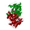
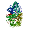
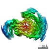
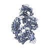
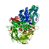
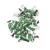
| Title | Crystal Structure of the Chorismate Synthase from Campylobacter jejuni, Northeast Structural Genomics Target BR19 | ||||||
 Components Components | Chorismate synthase | ||||||
 Keywords Keywords | LYASE / Structural Genomics / Bifunctional alpha/beta tetrameric protein / PSI / Protein Structure Initiative / Northeast Structural Genomics Consortium / NESG | ||||||
| Function / homology |  Function and homology information Function and homology informationchorismate synthase / chorismate synthase activity / chorismate biosynthetic process / aromatic amino acid family biosynthetic process Similarity search - Function | ||||||
| Biological species |   Campylobacter jejuni (Campylobacter) Campylobacter jejuni (Campylobacter) | ||||||
| Method |  X-RAY DIFFRACTION / SYNCHROTRON / SAD / Resolution: 2.8 Å X-RAY DIFFRACTION / SYNCHROTRON / SAD / Resolution: 2.8 Å | ||||||
 Authors Authors | Forouhar, F. / Lee, I. / Vorobiev, S.M. / Xiao, R. / Acton, T.B. / Montelione, G.T. / Tong, L. / Hunt, J.F. / Northeast Structural Genomics Consortium (NESG) | ||||||
 Citation Citation | Journal: To be Published Title: Crystal Structure of the Chorismate Synthase from Campylobacter jejuni, Northeast Structural Genomics Target BR19 Authors: Forouhar, F. / Lee, I. / Vorobiev, S.M. / Xiao, R. / Acton, T.B. / Montelione, G.T. / Tong, L. / Hunt, J.F. | ||||||
| History |
|
- Structure visualization
| Structure viewer | Molecule: MolmilJmol/JSmol |
|---|
- Downloads & links
-Download
| PDBx/mmCIF format | 1sq1.cif.gz | 63.9 KB | Display | PDBx/mmCIF format |
|---|---|---|---|---|
| PDB format | pdb1sq1.ent.gz | 51 KB | Display | PDB format |
| PDBx/mmJSON format | 1sq1.json.gz | Tree view | PDBx/mmJSON format | |
| Others |  Other downloads Other downloads |
-Validation report
| Summary document | 1sq1_validation.pdf.gz | 437.1 KB | Display | wwPDB validaton report |
|---|---|---|---|---|
| Full document | 1sq1_full_validation.pdf.gz | 445 KB | Display | |
| Data in XML | 1sq1_validation.xml.gz | 13.1 KB | Display | |
| Data in CIF | 1sq1_validation.cif.gz | 16.6 KB | Display | |
| Arichive directory | https://data.pdbj.org/pub/pdb/validation_reports/sq/1sq1ftp://data.pdbj.org/pub/pdb/validation_reports/sq/1sq1 | HTTPS FTP |
-Related structure data
| Similar structure data | |
|---|---|
| Other databases |

-Links
 PDBj
PDBj- Assembly
| Deposited unit | 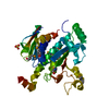
| ||||||||
|---|---|---|---|---|---|---|---|---|---|
| 1 | 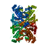
| ||||||||
| Unit cell |
|
-Components
| #1: Protein | Mass: 40705.129 Da / Num. of mol.: 1 Source method: isolated from a genetically manipulated source Source: (gene. exp.) Campylobacter jejuni (Campylobacter) / Strain: NCTC 11168 / Gene: AROC OR CJ1634 / Plasmid: pET21 / Production host: | ||
|---|---|---|---|
| #2: Chemical |   Mass: 96.063 Da / Num. of mol.: 2 / Source method: obtained synthetically / Formula: SO4 Mass: 96.063 Da / Num. of mol.: 2 / Source method: obtained synthetically / Formula: SO4#3: Water | ChemComp-HOH / |  Mass: 18.015 Da / Num. of mol.: 7 / Source method: isolated from a natural source / Formula: H2O Mass: 18.015 Da / Num. of mol.: 7 / Source method: isolated from a natural source / Formula: H2O |
-Experimental details
-Experiment
| Experiment | Method: X-RAY DIFFRACTION / Number of used crystals: 1 |
|---|
- Sample preparation
| Crystal | Density Matthews: 3.81 Å3/Da / Density % sol: 67.43 % |
|---|---|
| Crystal grow | Temperature: 277 K / Method: vapor diffusion, hanging drop Details: Protein solution: 10mM Tris (pH 7.5), 5mM DTT, 100mM NaCl. Well solution: 50mM NaAcetate (pH 4.6), 1M ammonium sulfate, 10mM DTT, 25mM octanoylsucrose, VAPOR DIFFUSION, HANGING DROP, temperature 277K |
-Data collection
| Diffraction | Mean temperature: 100 K |
|---|---|
| Diffraction source | Source: SYNCHROTRON / Site: NSLS  / Beamline: X4A / Wavelength: 0.97914 Å / Beamline: X4A / Wavelength: 0.97914 Å |
| Detector | Type: ADSC QUANTUM 4 / Detector: CCD / Date: Jan 30, 2004 / Details: mirrors |
| Radiation | Protocol: SINGLE WAVELENGTH / Monochromatic (M) / Laue (L): M / Scattering type: x-ray |
| Radiation wavelength | Wavelength: 0.97914 Å / Relative weight: 1 |
| Reflection | Resolution: 2.8→29.6 Å / Num. obs: 21612 / % possible obs: 98.9 % / Observed criterion σ(F): 2 / Observed criterion σ(I): 2 / Redundancy: 6.6 % / Biso Wilson estimate: -1.9 Å2 / Rmerge(I) obs: 0.061 / Rsym value: 0.062 / Net I/σ(I): 22.4 |
| Reflection shell | Resolution: 2.8→2.9 Å / Redundancy: 5.1 % / Rmerge(I) obs: 0.188 / Mean I/σ(I) obs: 6 / Num. unique all: 2148 / Rsym value: 0.19 / % possible all: 98.3 |
- Processing
| Software |
| |||||||||||||||||||||||||
|---|---|---|---|---|---|---|---|---|---|---|---|---|---|---|---|---|---|---|---|---|---|---|---|---|---|---|
| Refinement | Method to determine structure: SAD / Resolution: 2.8→29.6 Å / Isotropic thermal model: Overall anisotropic / Cross valid method: THROUGHOUT / σ(F): 2 / σ(I): 2 / Stereochemistry target values: Engh & Huber / Details: Used weighted full matrix least squares procedure.
| |||||||||||||||||||||||||
| Displacement parameters | Biso mean: 69.1 Å2
| |||||||||||||||||||||||||
| Refine analyze |
| |||||||||||||||||||||||||
| Refinement step | Cycle: LAST / Resolution: 2.8→29.6 Å
| |||||||||||||||||||||||||
| Refine LS restraints |
| |||||||||||||||||||||||||
| LS refinement shell | Resolution: 2.8→2.98 Å / Rfactor Rfree error: 0.024
|
