 Movie
Movie Controller


+ Open data

- Basic information
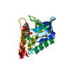
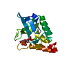
| Entry | Database: PDB / ID: 1snq | ||||||
|---|---|---|---|---|---|---|---|
| Title | PROTEIN STABILITY IN STAPHYLOCOCCAL NUCLEASE | ||||||
 Components Components | STAPHYLOCOCCAL NUCLEASE Micrococcal nuclease Micrococcal nuclease | ||||||
 Keywords Keywords | HYDROLASE / NUCLEASE / ENDONUCLEASE / CALCIUM | ||||||
| Function / homology |  Function and homology information Function and homology informationendonuclease activity, active with either ribo- or deoxyribonucleic acids and producing 3'-phosphomonoesters / micrococcal nuclease / nucleic acid binding / extracellular region / membrane / metal ion bindingSimilarity search - Function | ||||||
| Biological species |   Staphylococcus aureus (bacteria) Staphylococcus aureus (bacteria) | ||||||
| Method | X-RAY DIFFRACTION / Resolution: 1.7 Å | ||||||
 Authors Authors | Truckses, D.M. / Somoza, J.R. / Markley, J.L. | ||||||
 Citation Citation | Journal: Protein Sci. / Year: 1996 Title: Coupling between trans/cis proline isomerization and protein stability in staphylococcal nuclease. Authors: Truckses, D.M. / Somoza, J.R. / Prehoda, K.E. / Miller, S.C. / Markley, J.L. #1: Journal: Biochemistry / Year: 1994Title: Engineering Alternative Beta-Turn Types in Staphylococcal Nuclease Authors: Hynes, T.R. / Hodel, A. / Fox, R.O. #2: Journal: Proteins / Year: 1991Title: The Crystal Structure of Staphylococcal Nuclease Refined at 1.7 A Resolution Authors: Hynes, T.R. / Fox, R.O. #3: Journal: Biochemistry / Year: 1990Title: Coupling between Local Structure and Global Stability of a Protein: Mutants of Staphylococcal Nuclease Authors: Alexandrescu, A.T. / Hinck, A.P. / Markley, J.L. | ||||||
| History |
|
- Structure visualization
| Structure viewer | Molecule: MolmilJmol/JSmol |
|---|
- Downloads & links
-Download
| PDBx/mmCIF format | 1snq.cif.gz | 37.6 KB | Display | PDBx/mmCIF format |
|---|---|---|---|---|
| PDB format | pdb1snq.ent.gz | 25.9 KB | Display | PDB format |
| PDBx/mmJSON format | 1snq.json.gz | Tree view | PDBx/mmJSON format | |
| Others |  Other downloads Other downloads |
-Validation report
| Arichive directory | https://data.pdbj.org/pub/pdb/validation_reports/sn/1snqftp://data.pdbj.org/pub/pdb/validation_reports/sn/1snq | HTTPS FTP |
|---|
-Related structure data


-Links
 PDBj
PDBj- Assembly
| Deposited unit | 
| ||||||||
|---|---|---|---|---|---|---|---|---|---|
| 1 |
| ||||||||
| Unit cell |
|
-Components
| #1: Protein | Micrococcal nuclease / MICROCOCCAL NUCLEASE Mass: 16738.215 Da / Num. of mol.: 1 / Mutation: P47G, P117G Source method: isolated from a genetically manipulated source Details: THIS STAPH NUCLEASE IS FROM THE V8 STRAIN, THEREFORE RESIDUE 124 IS LEU, NOT HIS AS IN THE FOGGI STRAIN Source: (gene. exp.) Staphylococcus aureus (bacteria) / Strain: V8 / Description: PET SYSTEM / Gene: NUC / Plasmid: PET3A / Gene (production host): NUC / Production host: Escherichia coli (E. coli) / Strain (production host): PET / References: UniProt: P00644, micrococcal nuclease |
|---|---|
| #2: Water | ChemComp-HOH / Water Mass: 18.015 Da / Num. of mol.: 25 / Source method: isolated from a natural source / Formula: H2O Mass: 18.015 Da / Num. of mol.: 25 / Source method: isolated from a natural source / Formula: H2O |
-Experimental details
-Experiment
| Experiment | Method: X-RAY DIFFRACTION |
|---|
- Sample preparation
| Crystal | Density Matthews: 2.29 Å3/Da / Density % sol: 46.38 % | ||||||||||||||||||||||||||||||||||||||||||||||||
|---|---|---|---|---|---|---|---|---|---|---|---|---|---|---|---|---|---|---|---|---|---|---|---|---|---|---|---|---|---|---|---|---|---|---|---|---|---|---|---|---|---|---|---|---|---|---|---|---|---|
| Crystal grow | pH: 8.15 Details: 10.5 MM POTASSIUM PHOSPHATE, 20% - 30% 2-METHYL-2,4-PENTANEDIOL, pH 8.15 | ||||||||||||||||||||||||||||||||||||||||||||||||
| Crystal grow | *PLUS Temperature: 4 ℃ / Method: vapor diffusionDetails: Loll, P.J., (1989) Proteins: Struct.,Funct., Genet., 5, 183. | ||||||||||||||||||||||||||||||||||||||||||||||||
| Components of the solutions | *PLUS
|
-Data collection
| Diffraction | Mean temperature: 277 K |
|---|---|
| Diffraction source | Wavelength: 1.5418 |
| Detector | Type: RIGAKU RAXIS IIC / Detector: IMAGE PLATE / Date: Feb 1, 1993 |
| Radiation | Monochromatic (M) / Laue (L): M / Scattering type: x-ray |
| Radiation wavelength | Wavelength: 1.5418 Å / Relative weight: 1 |
| Reflection | Num. obs: 10604 / % possible obs: 94.5 % / Observed criterion σ(I): 0 / Rmerge(I) obs: 0.0474 |
| Reflection | *PLUS Highest resolution: 1.95 Å / Lowest resolution: 9999 Å / Num. measured all: 30773 |
| Reflection shell | *PLUS Highest resolution: 1.95 Å / Lowest resolution: 2 Å / % possible obs: 90 % |
- Processing
| Software |
| |||||||||||||||
|---|---|---|---|---|---|---|---|---|---|---|---|---|---|---|---|---|
| Refinement | Resolution: 1.7→6 Å / σ(F): 2
| |||||||||||||||
| Displacement parameters | Biso mean: 49.9 Å2 | |||||||||||||||
| Refinement step | Cycle: LAST / Resolution: 1.7→6 Å
| |||||||||||||||
| Software | *PLUS Name: X-PLOR / Classification: refinement | |||||||||||||||
| Refinement | *PLUS Num. reflection all: 10604 / Num. reflection obs: 9523 / Highest resolution: 1.95 Å | |||||||||||||||
| Solvent computation | *PLUS | |||||||||||||||
| Displacement parameters | *PLUS | |||||||||||||||
| Refine LS restraints | *PLUS
|
