 Movie
Movie Controller

[English] 日本語
 Yorodumi
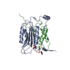
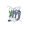
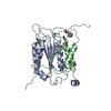
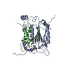
- PDB-1sbk: X-RAY STRUCTURE OF YDII_ECOLI NORTHEAST STRUCTURAL GENOMICS CONSO... -
+ Open data

- Basic information
| Entry | Database: PDB / ID: 1sbk | ||||||
|---|---|---|---|---|---|---|---|
| Title | X-RAY STRUCTURE OF YDII_ECOLI NORTHEAST STRUCTURAL GENOMICS CONSORTIUM TARGET ER29. | ||||||
 Components Components | Hypothetical protein ydiI Hypothesis Hypothesis | ||||||
 Keywords Keywords | STRUCTURAL GENOMICS / UNKNOWN FUNCTION / er29 / YDII / PSI / Protein Structure Initiative / Northeast Structural Genomics Consortium / NESG | ||||||
| Function / homology |  Function and homology information1,4-dihydroxy-2-naphthoyl-CoA hydrolase / 1,4-dihydroxy-2-naphthoyl-CoA thioesterase activity / acyl-CoA hydrolase activity / menaquinone biosynthetic process / hydrolase activity / cytosol Function and homology information1,4-dihydroxy-2-naphthoyl-CoA hydrolase / 1,4-dihydroxy-2-naphthoyl-CoA thioesterase activity / acyl-CoA hydrolase activity / menaquinone biosynthetic process / hydrolase activity / cytosolSimilarity search - Function | ||||||
| Biological species |  Escherichia coli (E. coli) Escherichia coli (E. coli) | ||||||
| Method | X-RAY DIFFRACTION / SYNCHROTRON / MOLECULAR REPLACEMENT / Resolution: 2 Å | ||||||
 Authors Authors | Kuzin, A.P. / Edstrom, W. / Vorobiev, S.M. / Lee, I. / Forouhar, F. / Ma, L. / Chiang, Y. / Rong, X. / Acton, T.B. / Montelione, G.T. ...Kuzin, A.P. / Edstrom, W. / Vorobiev, S.M. / Lee, I. / Forouhar, F. / Ma, L. / Chiang, Y. / Rong, X. / Acton, T.B. / Montelione, G.T. / Hunt, J.F. / Tong, L. / Northeast Structural Genomics Consortium (NESG) | ||||||
 Citation Citation | Journal: To be Published Title: X-ray Structure of YDII_ECOLI Northeast Structural Genomics Consortium Target ER29 Authors: Kuzin, A.P. / Edstrom, W. / Vorobiev, S.M. / Lee, I. / Forouhar, F. / Ma, L. / Chiang, Y. / Rong, X. / Acton, T.B. / Montelione, G.T. / Hunt, J.F. / Tong, L. | ||||||
| History |
|
- Structure visualization
| Structure viewer | Molecule: MolmilJmol/JSmol |
|---|
- Downloads & links
-Download
| PDBx/mmCIF format | 1sbk.cif.gz | 126.9 KB | Display | PDBx/mmCIF format |
|---|---|---|---|---|
| PDB format | pdb1sbk.ent.gz | 98.9 KB | Display | PDB format |
| PDBx/mmJSON format | 1sbk.json.gz | Tree view | PDBx/mmJSON format | |
| Others |  Other downloads Other downloads |
-Validation report
| Arichive directory | https://data.pdbj.org/pub/pdb/validation_reports/sb/1sbkftp://data.pdbj.org/pub/pdb/validation_reports/sb/1sbk | HTTPS FTP |
|---|
-Related structure data
| Related structure data |  1o0i S: Starting model for refinement |
|---|---|
| Similar structure data | |
| Other databases |


-Links
 PDBj
PDBj
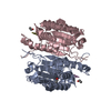
- Assembly
| Deposited unit | 
| ||||||||
|---|---|---|---|---|---|---|---|---|---|
| 1 |
| ||||||||
| Unit cell |
|
-Components
| #1: Protein | Hypothesis Mass: 16225.004 Da / Num. of mol.: 4 Source method: isolated from a genetically manipulated source Source: (gene. exp.) Escherichia coli (E. coli) / Gene: YDII, B1686 / Plasmid: pET21D / Species (production host): Escherichia coli / Production host: Escherichia coli BL21(DE3) (bacteria) / Strain (production host): BL21(DE3) / References: UniProt: P77781#2: Chemical | ChemComp-SO4 / Sulfate  Mass: 96.063 Da / Num. of mol.: 4 / Source method: obtained synthetically / Formula: SO4 Mass: 96.063 Da / Num. of mol.: 4 / Source method: obtained synthetically / Formula: SO4#3: Water | ChemComp-HOH / | Water Mass: 18.015 Da / Num. of mol.: 425 / Source method: isolated from a natural source / Formula: H2O Mass: 18.015 Da / Num. of mol.: 425 / Source method: isolated from a natural source / Formula: H2O |
|---|
-Experimental details
-Experiment
| Experiment | Method: X-RAY DIFFRACTION / Number of used crystals: 1 |
|---|
- Sample preparation
| Crystal | Density Matthews: 1.98 Å3/Da / Density % sol: 37.82 % |
|---|---|
| Crystal grow | Temperature: 303 K / Method: vapor diffusion, hanging drop / pH: 6 Details: 1.75M Ammonium sulfate, 0.1M NaCl, 10mM DTT, 50 mM Imidazole pH 6.0, 3% MPD, VAPOR DIFFUSION, HANGING DROP, temperature 303K |
-Data collection
| Diffraction | Mean temperature: 100 K |
|---|---|
| Diffraction source | Source: SYNCHROTRON / Site: NSLS  / Beamline: X4A / Wavelength: 0.97938 Å / Beamline: X4A / Wavelength: 0.97938 Å |
| Detector | Type: ADSC QUANTUM 4 / Detector: CCD / Date: Jan 31, 2004 |
| Radiation | Protocol: SINGLE WAVELENGTH / Monochromatic (M) / Laue (L): M / Scattering type: x-ray |
| Radiation wavelength | Wavelength: 0.97938 Å / Relative weight: 1 |
| Reflection | Resolution: 2→30 Å / Num. obs: 34984 / % possible obs: 99.5 % / Observed criterion σ(I): -3 / Redundancy: 4 % / Biso Wilson estimate: 19.7 Å2 / Rmerge(I) obs: 0.105 / Rsym value: 0.096 / Net I/σ(I): 13.3 |
| Reflection shell | Resolution: 2→2.07 Å / Rmerge(I) obs: 0.33 / Mean I/σ(I) obs: 4.1 / Num. unique all: 3466 / Rsym value: 0.27 / % possible all: 98.6 |
- Processing
| Software |
| |||||||||||||||||||||||||
|---|---|---|---|---|---|---|---|---|---|---|---|---|---|---|---|---|---|---|---|---|---|---|---|---|---|---|
| Refinement | Method to determine structure: MOLECULAR REPLACEMENT Starting model: PDB ENTRY 1O0I 1o0i Resolution: 2→28.97 Å / Rfactor Rfree error: 0.007 / Data cutoff high absF: 174379.22 / Data cutoff low absF: 0 / Isotropic thermal model: RESTRAINED / Cross valid method: THROUGHOUT / σ(F): 0 / Stereochemistry target values: Engh & Huber
| |||||||||||||||||||||||||
| Solvent computation | Solvent model: FLAT MODEL / Bsol: 61.3954 Å2 / ksol: 0.350749 e/Å3 | |||||||||||||||||||||||||
| Displacement parameters | Biso mean: 19.8 Å2
| |||||||||||||||||||||||||
| Refine analyze |
| |||||||||||||||||||||||||
| Refinement step | Cycle: LAST / Resolution: 2→28.97 Å
| |||||||||||||||||||||||||
| Refine LS restraints |
| |||||||||||||||||||||||||
| LS refinement shell | Resolution: 2→2.13 Å / Rfactor Rfree error: 0.018 / Total num. of bins used: 6
| |||||||||||||||||||||||||
| Xplor file |
|
