 Movie
Movie Controller


+ Open data

- Basic information
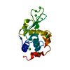
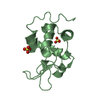
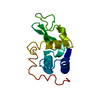
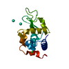
| Entry | Database: PDB / ID: 1qsw | ||||||
|---|---|---|---|---|---|---|---|
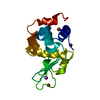
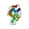
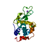
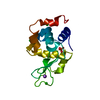
| Title | CRYSTAL STRUCTURE ANALYSIS OF A HUMAN LYSOZYME MUTANT W64C C65A | ||||||
 Components Components | HUMAN LYSOZYME MUTANT | ||||||
 Keywords Keywords |  HYDROLASE HYDROLASE | ||||||
| Function / homology |  Function and homology information Function and homology informationantimicrobial humoral response / Antimicrobial peptides / metabolic process / specific granule lumen / azurophil granule lumen / tertiary granule lumen / lysozyme / lysozyme activity / killing of cells of another organism / defense response to Gram-negative bacterium ...antimicrobial humoral response / Antimicrobial peptides / metabolic process / specific granule lumen / azurophil granule lumen / tertiary granule lumen / lysozyme / lysozyme activity / killing of cells of another organism / defense response to Gram-negative bacterium / defense response to Gram-positive bacterium / defense response to bacterium / inflammatory response / Amyloid fiber formation / Neutrophil degranulation / extracellular space / extracellular exosome / extracellular region / identical protein bindingSimilarity search - Function | ||||||
| Biological species |  Homo sapiens (human) Homo sapiens (human) | ||||||
| Method | X-RAY DIFFRACTION / Resolution: 1.85 Å | ||||||
 Authors Authors | Inaka, K. / Kanaya, E. / Kikuchi, M. / Miki, K. | ||||||
 Citation Citation | Journal: Proteins / Year: 2001 Title: Crystal structure of a mutant human lysozyme with a substituted disulfide bond. Authors: Inaka, K. / Kanaya, E. / Kikuchi, M. / Miki, K. | ||||||
| History |
|
- Structure visualization
| Structure viewer | Molecule: MolmilJmol/JSmol |
|---|
- Downloads & links
-Download
| PDBx/mmCIF format | 1qsw.cif.gz | 110.8 KB | Display | PDBx/mmCIF format |
|---|---|---|---|---|
| PDB format | pdb1qsw.ent.gz | 91.7 KB | Display | PDB format |
| PDBx/mmJSON format | 1qsw.json.gz | Tree view | PDBx/mmJSON format | |
| Others |  Other downloads Other downloads |
-Validation report
| Arichive directory | https://data.pdbj.org/pub/pdb/validation_reports/qs/1qswftp://data.pdbj.org/pub/pdb/validation_reports/qs/1qsw | HTTPS FTP |
|---|
-Related structure data
| Similar structure data |
|---|

-Links
 PDBj
PDBj

- Assembly
| Deposited unit | 
| ||||||||
|---|---|---|---|---|---|---|---|---|---|
| 1 | 
| ||||||||
| 2 | 
| ||||||||
| 3 | 
| ||||||||
| 4 | 
| ||||||||
| Unit cell |
|
-Components
| #1: Protein | Mass: 14605.561 Da / Num. of mol.: 4 / Mutation: W64C, C65A / Source method: isolated from a natural source / Source: (natural) Homo sapiens (human) / References: UniProt: P61626#2: Water | ChemComp-HOH / | Water Mass: 18.015 Da / Num. of mol.: 232 / Source method: isolated from a natural source / Formula: H2O Mass: 18.015 Da / Num. of mol.: 232 / Source method: isolated from a natural source / Formula: H2O |
|---|
-Experimental details
-Experiment
| Experiment | Method: X-RAY DIFFRACTION / Number of used crystals: 1 |
|---|
- Sample preparation
| Crystal | Density Matthews: 2.47 Å3/Da / Density % sol: 50.19 % | ||||||||||||||||||||||||
|---|---|---|---|---|---|---|---|---|---|---|---|---|---|---|---|---|---|---|---|---|---|---|---|---|---|
| Crystal grow | Temperature: 298 K / Method: vapor diffusion, hanging drop / pH: 6 Details: SODIUM PHOSPHATE, NACL, pH 6.0, VAPOR DIFFUSION, HANGING DROP, temperature 25K | ||||||||||||||||||||||||
| Crystal grow | *PLUS Method: unknown | ||||||||||||||||||||||||
| Components of the solutions | *PLUS
|
-Data collection
| Diffraction | Mean temperature: 298 K |
|---|---|
| Diffraction source | Source: ROTATING ANODE / Type: MACSCIENCE / Wavelength: 1.5418 |
| Detector | Type: MACSCIENCE DIP100S / Detector: IMAGE PLATE / Date: Feb 12, 1991 |
| Radiation | Protocol: SINGLE WAVELENGTH / Monochromatic (M) / Laue (L): M / Scattering type: x-ray |
| Radiation wavelength | Wavelength: 1.5418 Å / Relative weight: 1 |
| Reflection | Resolution: 1.85→16 Å / Num. all: 76633 / Num. obs: 43715 / % possible obs: 80.4 % / Observed criterion σ(F): 3 / Observed criterion σ(I): 3 / Rmerge(I) obs: 0.066 |
| Reflection shell | Resolution: 1.85→1.94 Å / % possible all: 63.2 |
| Reflection | *PLUS Lowest resolution: 30 Å / Num. measured all: 76633 / Rmerge(I) obs: 0.066 |
| Reflection shell | *PLUS % possible obs: 64.3 % / Num. unique obs: 3092 / Num. measured obs: 6244 / Rmerge(I) obs: 0.163 / Mean I/σ(I) obs: 2.1 |
- Processing
| Software |
| |||||||||||||||||||||||||||||||||||||||||||||||||||||||||||||||
|---|---|---|---|---|---|---|---|---|---|---|---|---|---|---|---|---|---|---|---|---|---|---|---|---|---|---|---|---|---|---|---|---|---|---|---|---|---|---|---|---|---|---|---|---|---|---|---|---|---|---|---|---|---|---|---|---|---|---|---|---|---|---|---|---|
| Refinement | Resolution: 1.85→10 Å / σ(F): 3 / σ(I): 3 / Stereochemistry target values: ENGH & HUBER
| |||||||||||||||||||||||||||||||||||||||||||||||||||||||||||||||
| Refinement step | Cycle: LAST / Resolution: 1.85→10 Å
| |||||||||||||||||||||||||||||||||||||||||||||||||||||||||||||||
| Refine LS restraints |
| |||||||||||||||||||||||||||||||||||||||||||||||||||||||||||||||
| Software | *PLUS Name: PROLSQ / Classification: refinement | |||||||||||||||||||||||||||||||||||||||||||||||||||||||||||||||
| Refinement | *PLUS σ(F): 3 / Rfactor obs: 0.181 | |||||||||||||||||||||||||||||||||||||||||||||||||||||||||||||||
| Solvent computation | *PLUS | |||||||||||||||||||||||||||||||||||||||||||||||||||||||||||||||
| Displacement parameters | *PLUS |
