 Movie
Movie Controller

[English] 日本語
 Yorodumi
- PDB-1qin: HUMAN GLYOXALASE I COMPLEXED WITH S-(N-HYDROXY-N-P-IODOPHENYLCARB... -
+ Open data

- Basic information
| Entry | Database: PDB / ID: 1qin | ||||||
|---|---|---|---|---|---|---|---|
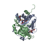
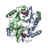
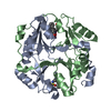
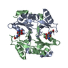
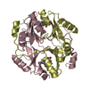
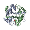
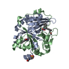
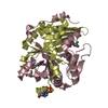
| Title | HUMAN GLYOXALASE I COMPLEXED WITH S-(N-HYDROXY-N-P-IODOPHENYLCARBAMOYL) GLUTATHIONE | ||||||
 Components Components | PROTEIN (LACTOYLGLUTATHIONE LYASE) | ||||||
 Keywords Keywords | LYASE / LACTOYLGLUTATHIONE LYASE / GLYOXALASE I | ||||||
| Function / homology |  Function and homology information Function and homology informationlactoylglutathione lyase / lactoylglutathione lyase activity / methylglyoxal metabolic process / Pyruvate metabolism / glutathione metabolic process / osteoclast differentiation / carbohydrate metabolic process / regulation of transcription by RNA polymerase II / negative regulation of apoptotic process / extracellular exosome ...lactoylglutathione lyase / lactoylglutathione lyase activity / methylglyoxal metabolic process / Pyruvate metabolism / glutathione metabolic process / osteoclast differentiation / carbohydrate metabolic process / regulation of transcription by RNA polymerase II / negative regulation of apoptotic process / extracellular exosome / zinc ion binding / nucleoplasm / plasma membrane / cytoplasm / cytosol Similarity search - Function | ||||||
| Biological species |  Homo sapiens (human) Homo sapiens (human) | ||||||
| Method |  X-RAY DIFFRACTION / OTHER / Resolution: 2 Å X-RAY DIFFRACTION / OTHER / Resolution: 2 Å | ||||||
 Authors Authors | Cameron, A.D. / Ridderstrom, M. / Olin, B. / Mannervik, B. | ||||||
 Citation Citation | Journal: Biochemistry / Year: 1999 Title: Reaction mechanism of glyoxalase I explored by an X-ray crystallographic analysis of the human enzyme in complex with a transition state analogue. Authors: Cameron, A.D. / Ridderstrom, M. / Olin, B. / Kavarana, M.J. / Creighton, D.J. / Mannervik, B. | ||||||
| History |
|
- Structure visualization
| Structure viewer | Molecule: MolmilJmol/JSmol |
|---|
- Downloads & links
-Download
| PDBx/mmCIF format | 1qin.cif.gz | 88.9 KB | Display | PDBx/mmCIF format |
|---|---|---|---|---|
| PDB format | pdb1qin.ent.gz | 66.9 KB | Display | PDB format |
| PDBx/mmJSON format | 1qin.json.gz | Tree view | PDBx/mmJSON format | |
| Others |  Other downloads Other downloads |
-Validation report
| Arichive directory | https://data.pdbj.org/pub/pdb/validation_reports/qi/1qinftp://data.pdbj.org/pub/pdb/validation_reports/qi/1qin | HTTPS FTP |
|---|
-Related structure data
-Links
 PDBj
PDBj- Assembly
| Deposited unit | 
| ||||||||
|---|---|---|---|---|---|---|---|---|---|
| 1 |
| ||||||||
| Unit cell |
| ||||||||
| Noncrystallographic symmetry (NCS) | NCS oper: (Code: given Matrix: (-0.9898, -0.12358, 0.07082), Vector: |
-Components
| #1: Protein | Mass: 20672.520 Da / Num. of mol.: 2 Source method: isolated from a genetically manipulated source Source: (gene. exp.) Homo sapiens (human) / Description: HETEROLOGOUSLY EXPRESSED / Plasmid: PKK223-3 / Production host:  #2: Chemical |   Mass: 65.409 Da / Num. of mol.: 2 / Source method: obtained synthetically / Formula: Zn Mass: 65.409 Da / Num. of mol.: 2 / Source method: obtained synthetically / Formula: Zn#3: Chemical |   Mass: 570.356 Da / Num. of mol.: 2 / Source method: obtained synthetically / Formula: C17H23IN4O8S Mass: 570.356 Da / Num. of mol.: 2 / Source method: obtained synthetically / Formula: C17H23IN4O8S#4: Water | ChemComp-HOH / |  Mass: 18.015 Da / Num. of mol.: 191 / Source method: isolated from a natural source / Formula: H2O Mass: 18.015 Da / Num. of mol.: 191 / Source method: isolated from a natural source / Formula: H2OSequence details | THE REFERENCE FOR THE SEQUENCE: M.RIDDERSTRO | |
|---|
-Experimental details
-Experiment
| Experiment | Method: X-RAY DIFFRACTION / Number of used crystals: 1 |
|---|
- Sample preparation
| Crystal | Density Matthews: 2.9 Å3/Da / Density % sol: 49 % | ||||||||||||||||||||||||||||||||||||||||||||||||||||||||||||
|---|---|---|---|---|---|---|---|---|---|---|---|---|---|---|---|---|---|---|---|---|---|---|---|---|---|---|---|---|---|---|---|---|---|---|---|---|---|---|---|---|---|---|---|---|---|---|---|---|---|---|---|---|---|---|---|---|---|---|---|---|---|
| Crystal grow | pH: 5.8 Details: PROTEIN WAS CRYSTALLISED BY EQILLABRATION AGAINST PEG 2000 MONOMETHLY ETHER, 50 MM MES PH 5.8, 0.1M NACL. HIPC-GSH WAS PRESENT IN THE PROTEIN SOLUTION | ||||||||||||||||||||||||||||||||||||||||||||||||||||||||||||
| Crystal | *PLUS | ||||||||||||||||||||||||||||||||||||||||||||||||||||||||||||
| Crystal grow | *PLUS Temperature: 15 ℃ / Method: vapor diffusion, hanging drop / Details: Cameron, A.D., (1997) EMBO J., 16, 3386. | ||||||||||||||||||||||||||||||||||||||||||||||||||||||||||||
| Components of the solutions | *PLUS
|
-Data collection
| Diffraction | Mean temperature: 263 K |
|---|---|
| Diffraction source | Source: ROTATING ANODE / Type: RIGAKU RU200 / Wavelength: 1.54 |
| Detector | Type: RIGAKU RAXIS II / Detector: IMAGE PLATE / Date: Nov 3, 1997 / Details: COLLIMATOR |
| Radiation | Monochromator: GRAPHITE / Protocol: SINGLE WAVELENGTH / Monochromatic (M) / Laue (L): M / Scattering type: x-ray |
| Radiation wavelength | Wavelength: 1.54 Å / Relative weight: 1 |
| Reflection | Resolution: 2→29 Å / Num. obs: 28148 / % possible obs: 93.6 % / Redundancy: 1.8 % / Biso Wilson estimate: 23 Å2 / Rmerge(I) obs: 0.83 / Net I/σ(I): 9 |
| Reflection shell | Resolution: 2→2.11 Å / Redundancy: 1.7 % / Rmerge(I) obs: 0.194 / Mean I/σ(I) obs: 3.2 / % possible all: 93.5 |
| Reflection | *PLUS Num. measured all: 49286 |
| Reflection shell | *PLUS % possible obs: 93.5 % |
- Processing
| Software |
| ||||||||||||||||||||||||||||||||||||||||||||||||||||||||||||||||||||||||||||||||||||
|---|---|---|---|---|---|---|---|---|---|---|---|---|---|---|---|---|---|---|---|---|---|---|---|---|---|---|---|---|---|---|---|---|---|---|---|---|---|---|---|---|---|---|---|---|---|---|---|---|---|---|---|---|---|---|---|---|---|---|---|---|---|---|---|---|---|---|---|---|---|---|---|---|---|---|---|---|---|---|---|---|---|---|---|---|---|
| Refinement | Method to determine structure: OTHER / Resolution: 2→29 Å / SU B: 3.3 / SU ML: 0.09 / Cross valid method: THROUGHOUT / σ(F): 0 / ESU R: 0.17 / ESU R Free: 0.15
| ||||||||||||||||||||||||||||||||||||||||||||||||||||||||||||||||||||||||||||||||||||
| Displacement parameters | Biso mean: 30 Å2 | ||||||||||||||||||||||||||||||||||||||||||||||||||||||||||||||||||||||||||||||||||||
| Refinement step | Cycle: LAST / Resolution: 2→29 Å
| ||||||||||||||||||||||||||||||||||||||||||||||||||||||||||||||||||||||||||||||||||||
| Refine LS restraints |
| ||||||||||||||||||||||||||||||||||||||||||||||||||||||||||||||||||||||||||||||||||||
| Software | *PLUS Name: REFMAC / Classification: refinement | ||||||||||||||||||||||||||||||||||||||||||||||||||||||||||||||||||||||||||||||||||||
| Refinement | *PLUS Highest resolution: 2 Å / Lowest resolution: 29 Å / σ(F): 0 / % reflection Rfree: 5 % / Rfactor obs: 0.18 / Rfactor Rfree: 0.21 | ||||||||||||||||||||||||||||||||||||||||||||||||||||||||||||||||||||||||||||||||||||
| Solvent computation | *PLUS | ||||||||||||||||||||||||||||||||||||||||||||||||||||||||||||||||||||||||||||||||||||
| Displacement parameters | *PLUS Biso mean: 30 Å2 | ||||||||||||||||||||||||||||||||||||||||||||||||||||||||||||||||||||||||||||||||||||
| Refine LS restraints | *PLUS
|
