[English] 日本語
Yorodumi
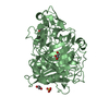
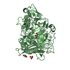
- PDB-1q9h: 3-Dimensional structure of native Cel7A from Talaromyces emersonii -

+
Open data


ID or keywords:

Loading...

-
Basic information

Entry
Database: PDB / ID: 1q9h
Title3-Dimensional structure of native Cel7A from Talaromyces emersonii
Componentscellobiohydrolase I catalytic domain
KeywordsHYDROLASE / cellobiohydrolase I catalytic domain
Function / homology
Function and homology information


Hydrolases; Glycosylases; Glycosidases, i.e. enzymes that hydrolyse O- and S-glycosyl compounds / cellulose catabolic process / hydrolase activity, hydrolyzing O-glycosyl compounds
Similarity search - Function
1,4-Beta-D-Glucan Cellobiohydrolase I; Chain A / Glycoside hydrolase, family 7, domain / Glycoside hydrolase, family 7 / Glycoside hydrolase family 7, catalytic domain superfamily / Glycosyl hydrolase family 7 / Distorted Sandwich / Concanavalin A-like lectin/glucanase domain superfamily / Mainly Beta
Similarity search - Domain/homology
Biological speciesTalaromyces emersonii (fungus)
MethodX-RAY DIFFRACTION / SYNCHROTRON / MOLECULAR REPLACEMENT / Resolution: 2.35 Å
AuthorsGrassick, A. / Thompson, R. / Murray, P.G. / Collins, C.M. / Byrnes, L. / Tuohy, M.G. / Birrane, G. / Higgins, T.M.
Citation
Journal: Eur.J.Biochem. / Year: 2004
Title: Three-dimensional structure of a thermostable native cellobiohydrolase, CBH IB, and molecular characterization of the cel7 gene from the filamentous fungus, Talaromyces emersonii
Authors: Grassick, A. / Murray, P.G. / Thompson, R. / Collins, C.M. / Byrnes, L. / Birrane, G. / Higgins, T.M. / Tuohy, M.G.
#1: Journal: Acta Crystallogr.,Sect.D / Year: 2003
Title: Crystallisation and preliminary crystallographic analysis of the catalytic domain Cellobiohydrolase IB from Talaromyces emersonii
Authors: Grassick, A. / Birrane, G. / Tuohy, M.G. / Murray, P.G. / Higgins, T.M.
History
DepositionAug 25, 2003Deposition site: RCSB / Processing site: RCSB
Revision 1.0Nov 9, 2004Provider: repository / Type: Initial release
Revision 1.1Apr 29, 2008Group: Version format compliance
Revision 1.2Jul 13, 2011Group: Non-polymer description / Version format compliance
Revision 2.0Dec 25, 2019Group: Data collection / Database references ...Data collection / Database references / Derived calculations / Polymer sequence
Category: diffrn_source / entity_poly ...diffrn_source / entity_poly / pdbx_struct_mod_residue / struct_conn / struct_ref_seq_dif
Item: _diffrn_source.pdbx_synchrotron_site / _entity_poly.pdbx_seq_one_letter_code_can ..._diffrn_source.pdbx_synchrotron_site / _entity_poly.pdbx_seq_one_letter_code_can / _pdbx_struct_mod_residue.parent_comp_id / _struct_conn.pdbx_leaving_atom_flag
Revision 3.0Jul 29, 2020Group: Atomic model / Data collection ...Atomic model / Data collection / Derived calculations / Structure summary
Category: atom_site / chem_comp ...atom_site / chem_comp / entity / pdbx_branch_scheme / pdbx_chem_comp_identifier / pdbx_entity_branch / pdbx_entity_branch_descriptor / pdbx_entity_branch_link / pdbx_entity_branch_list / pdbx_entity_nonpoly / pdbx_nonpoly_scheme / pdbx_struct_assembly_gen / struct_asym / struct_conn / struct_site / struct_site_gen
Item: _atom_site.auth_asym_id / _atom_site.auth_seq_id ..._atom_site.auth_asym_id / _atom_site.auth_seq_id / _atom_site.label_asym_id / _atom_site.label_entity_id / _chem_comp.name / _chem_comp.type / _pdbx_entity_nonpoly.entity_id / _pdbx_entity_nonpoly.name / _pdbx_struct_assembly_gen.asym_id_list / _struct_conn.pdbx_dist_value / _struct_conn.pdbx_leaving_atom_flag / _struct_conn.pdbx_role / _struct_conn.ptnr1_auth_asym_id / _struct_conn.ptnr1_auth_comp_id / _struct_conn.ptnr1_auth_seq_id / _struct_conn.ptnr1_label_asym_id / _struct_conn.ptnr1_label_atom_id / _struct_conn.ptnr1_label_comp_id / _struct_conn.ptnr1_label_seq_id / _struct_conn.ptnr2_auth_asym_id / _struct_conn.ptnr2_auth_comp_id / _struct_conn.ptnr2_auth_seq_id / _struct_conn.ptnr2_label_asym_id / _struct_conn.ptnr2_label_atom_id / _struct_conn.ptnr2_label_comp_id / _struct_conn.ptnr2_label_seq_id
Description: Carbohydrate remediation / Provider: repository / Type: Remediation

-
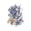
Structure visualization

Structure viewerMolecule:
MolmilJmol/JSmol

Downloads & links

-
Assembly

Deposited unit
A: cellobiohydrolase I catalytic domain
hetero molecules


Theoretical massNumber of molelcules
Total (without water)47,4953
Polymers46,8501
Non-polymers6462
Water4,576254
1


  • Idetical with deposited unit
  • defined by author
TypeNameSymmetry operationNumber
identity operation1_555x,y,z1
Unit cell
Length a, b, c (Å)74.395, 74.395, 177.046
Angle α, β, γ (deg.)90.00, 90.00, 90.00
Int Tables number92
Space group name H-MP41212

-
Components

#1: Protein cellobiohydrolase I catalytic domain


Mass: 46849.668 Da / Num. of mol.: 1 / Fragment: N-acetylglucosamine / Source method: isolated from a natural source / Details: Thermophilic Fungus / Source: (natural) Talaromyces emersonii (fungus) / Strain: CBS814.70
References: GenBank: 19568104, UniProt: Q8TFL9*PLUS, cellulose 1,4-beta-cellobiosidase (non-reducing end)
#2: Polysaccharide 2-acetamido-2-deoxy-beta-D-glucopyranose-(1-4)-2-acetamido-2-deoxy-beta-D-glucopyranose


Type: oligosaccharide / Mass: 424.401 Da / Num. of mol.: 1 / Fragment: water / Source method: isolated from a natural source
DescriptorTypeProgram
DGlcpNAcb1-4DGlcpNAcb1-Glycam Condensed SequenceGMML 1.0
WURCS=2.0/1,2,1/[a2122h-1b_1-5_2*NCC/3=O]/1-1/a4-b1WURCSPDB2Glycan 1.1.0
[]{[(4+1)][b-D-GlcpNAc]{[(4+1)][b-D-GlcpNAc]{}}}LINUCSPDB-CARE
#3: Sugar ChemComp-NAG / 2-acetamido-2-deoxy-beta-D-glucopyranose / N-Acetylglucosamine


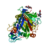
Type: D-saccharide, beta linking / Mass: 221.208 Da / Num. of mol.: 1 / Fragment: water / Source method: isolated from a natural source / Formula: C8H15NO6
IdentifierTypeProgram
DGlcpNAcbCONDENSED IUPAC CARBOHYDRATE SYMBOLGMML 1.0
N-acetyl-b-D-glucopyranosamineCOMMON NAMEGMML 1.0
b-D-GlcpNAcIUPAC CARBOHYDRATE SYMBOLPDB-CARE 1.0
GlcNAcSNFG CARBOHYDRATE SYMBOLGMML 1.0
#4: Water ChemComp-HOH / water / Water


Mass: 18.015 Da / Num. of mol.: 254 / Source method: isolated from a natural source / Formula: H2O

-
Experimental details

-
Experiment

ExperimentMethod: X-RAY DIFFRACTION / Number of used crystals: 1

-
Sample preparation

CrystalDensity Matthews: 2.61 Å3/Da / Density % sol: 52.95 %
Crystal growTemperature: 291 K / Method: vapor diffusion, hanging drop / pH: 8.5
Details: 2.5 M ammonium phosphate, 100 mM Tris-HCl, pH 8.5, VAPOR DIFFUSION, HANGING DROP, temperature 291K

-
Data collection

DiffractionMean temperature: 298 K
Diffraction sourceSource: SYNCHROTRON / Site: EMBL/DESY, HAMBURG / Beamline: BW7B / Wavelength: 0.8384 Å
DetectorType: MARRESEARCH / Detector: AREA DETECTOR / Date: Jan 1, 1996
RadiationMonochromator: Mirror / Protocol: SINGLE WAVELENGTH / Monochromatic (M) / Laue (L): M / Scattering type: x-ray
Radiation wavelengthWavelength: 0.8384 Å / Relative weight: 1
ReflectionResolution: 2.35→19.65 Å / Num. all: 21540 / Num. obs: 20984

-
Processing

Software
NameVersionClassification
REFMAC5.1.24refinement
DENZOdata reduction
SCALEPACKdata scaling
AMoREphasing
RefinementMethod to determine structure: MOLECULAR REPLACEMENT / Resolution: 2.35→19.65 Å / Cor.coef. Fo:Fc: 0.943 / Cor.coef. Fo:Fc free: 0.842 / SU B: 6.251 / SU ML: 0.147 / Cross valid method: THROUGHOUT / ESU R: 0.3 / ESU R Free: 0.232 / Stereochemistry target values: MAXIMUM LIKELIHOOD / Details: HYDROGENS HAVE BEEN ADDED IN THE RIDING POSITIONS
RfactorNum. reflection% reflectionSelection details
Rfree0.22934 1083 5.2 %RANDOM
Rwork0.16126 ---
obs0.16462 19901 97.66 %-
all-20984 --
Solvent computationIon probe radii: 0.8 Å / Shrinkage radii: 0.8 Å / VDW probe radii: 1.4 Å / Solvent model: BABINET MODEL WITH MASK
Displacement parametersBiso mean: 18.78 Å2
Baniso -1Baniso -2Baniso -3
1-0 Å20 Å20 Å2
2--0 Å20 Å2
3---0 Å2
Refinement stepCycle: LAST / Resolution: 2.35→19.65 Å
ProteinNucleic acidLigandSolventTotal
Num. atoms3208 0 42 254 3504
Refine LS restraints
Refine-IDTypeDev idealDev ideal targetNumber
X-RAY DIFFRACTIONr_bond_refined_d0.0120.0213341
X-RAY DIFFRACTIONr_bond_other_d0.0020.022718
X-RAY DIFFRACTIONr_angle_refined_deg1.4921.954574
X-RAY DIFFRACTIONr_angle_other_deg0.88436373
X-RAY DIFFRACTIONr_dihedral_angle_1_deg7.0095428
X-RAY DIFFRACTIONr_dihedral_angle_2_deg
X-RAY DIFFRACTIONr_dihedral_angle_3_deg
X-RAY DIFFRACTIONr_dihedral_angle_4_deg
X-RAY DIFFRACTIONr_chiral_restr0.090.2500
X-RAY DIFFRACTIONr_gen_planes_refined0.0050.023827
X-RAY DIFFRACTIONr_gen_planes_other0.0020.02637
X-RAY DIFFRACTIONr_nbd_refined0.2050.2626
X-RAY DIFFRACTIONr_nbd_other0.2430.23281
X-RAY DIFFRACTIONr_nbtor_refined
X-RAY DIFFRACTIONr_nbtor_other0.0870.21896
X-RAY DIFFRACTIONr_xyhbond_nbd_refined0.1690.2186
X-RAY DIFFRACTIONr_xyhbond_nbd_other
X-RAY DIFFRACTIONr_metal_ion_refined
X-RAY DIFFRACTIONr_metal_ion_other
X-RAY DIFFRACTIONr_symmetry_vdw_refined0.1920.26
X-RAY DIFFRACTIONr_symmetry_vdw_other0.1940.219
X-RAY DIFFRACTIONr_symmetry_hbond_refined0.170.213
X-RAY DIFFRACTIONr_symmetry_hbond_other
X-RAY DIFFRACTIONr_mcbond_it0.7271.52129
X-RAY DIFFRACTIONr_mcbond_other
X-RAY DIFFRACTIONr_mcangle_it1.35823426
X-RAY DIFFRACTIONr_scbond_it1.88731212
X-RAY DIFFRACTIONr_scangle_it3.1644.51148
X-RAY DIFFRACTIONr_rigid_bond_restr
X-RAY DIFFRACTIONr_sphericity_free
X-RAY DIFFRACTIONr_sphericity_bonded
LS refinement shellResolution: 2.35→2.41 Å / Total num. of bins used: 20 /
RfactorNum. reflection
Rfree0.291 91
Rwork0.213 1441

+
About Yorodumi

-
News

-
Feb 9, 2022. New format data for meta-information of EMDB entries

New format data for meta-information of EMDB entries

  • Version 3 of the EMDB header file is now the official format.
  • The previous official version 1.9 will be removed from the archive.

Related info.:EMDB header

External links:wwPDB to switch to version 3 of the EMDB data model

-
Aug 12, 2020. Covid-19 info

Covid-19 info

URL: https://pdbj.org/emnavi/covid19.php

New page: Covid-19 featured information page in EM Navigator.

Related info.:Covid-19 info / Mar 5, 2020. Novel coronavirus structure data

+
Mar 5, 2020. Novel coronavirus structure data

Novel coronavirus structure data

Related info.:Yorodumi Speices / Aug 12, 2020. Covid-19 info

External links:COVID-19 featured content - PDBj / Molecule of the Month (242):Coronavirus Proteases

+
Jan 31, 2019. EMDB accession codes are about to change! (news from PDBe EMDB page)

EMDB accession codes are about to change! (news from PDBe EMDB page)

  • The allocation of 4 digits for EMDB accession codes will soon come to an end. Whilst these codes will remain in use, new EMDB accession codes will include an additional digit and will expand incrementally as the available range of codes is exhausted. The current 4-digit format prefixed with “EMD-” (i.e. EMD-XXXX) will advance to a 5-digit format (i.e. EMD-XXXXX), and so on. It is currently estimated that the 4-digit codes will be depleted around Spring 2019, at which point the 5-digit format will come into force.
  • The EM Navigator/Yorodumi systems omit the EMD- prefix.

Related info.:Q: What is EMD? / ID/Accession-code notation in Yorodumi/EM Navigator

External links:EMDB Accession Codes are Changing Soon! / Contact to PDBj

+
Jul 12, 2017. Major update of PDB

Major update of PDB

  • wwPDB released updated PDB data conforming to the new PDBx/mmCIF dictionary.
  • This is a major update changing the version number from 4 to 5, and with Remediation, in which all the entries are updated.
  • In this update, many items about electron microscopy experimental information are reorganized (e.g. em_software).
  • Now, EM Navigator and Yorodumi are based on the updated data.

External links:wwPDB Remediation / Enriched Model Files Conforming to OneDep Data Standards Now Available in the PDB FTP Archive

-
Yorodumi

Thousand views of thousand structures

  • Yorodumi is a browser for structure data from EMDB, PDB, SASBDB, etc.
  • This page is also the successor to EM Navigator detail page, and also detail information page/front-end page for Omokage search.
  • The word "yorodu" (or yorozu) is an old Japanese word meaning "ten thousand". "mi" (miru) is to see.

Related info.:EMDB / PDB / SASBDB / Comparison of 3 databanks / Yorodumi Search / Aug 31, 2016. New EM Navigator & Yorodumi / Yorodumi Papers / Jmol/JSmol / Function and homology information / Changes in new EM Navigator and Yorodumi

Read more