 Movie
Movie Controller


+ Open data

- Basic information
| Entry | Database: PDB / ID: 1pd3 | ||||||
|---|---|---|---|---|---|---|---|
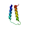
| Title | Influenza A NEP M1-binding domain | ||||||
 Components Components | Nonstructural protein NS2 | ||||||
 Keywords Keywords | UNKNOWN FUNCTION / influenza virus A / NEP/NS2 | ||||||
| Function / homology |  Function and homology information Function and homology informationuncoating of virus / exit of virus from host cell nucleus through nuclear pore / intracellular transport of virus / Influenza Infection / Fusion of the Influenza Virion to the Host Cell Endosome / Release /  Budding / Packaging of Eight RNA Segments / Uncoating of the Influenza Virion / Entry of Influenza Virion into Host Cell via Endocytosis ...uncoating of virus / exit of virus from host cell nucleus through nuclear pore / intracellular transport of virus / Influenza Infection / Fusion of the Influenza Virion to the Host Cell Endosome / Release / Budding / Packaging of Eight RNA Segments / Uncoating of the Influenza Virion / Entry of Influenza Virion into Host Cell via Endocytosis / viral genome packaging / Viral RNP Complexes in the Host Cell Nucleus / virion component => GO:0044423 / NEP/NS2 Interacts with the Cellular Export Machinery / Viral mRNA Translation / viral budding from plasma membrane / endosome lumen / endocytic vesicle membrane / receptor-mediated endocytosis of virus by host cell / fusion of virus membrane with host plasma membrane / host cell nucleus / virion attachment to host cell / extracellular region / nucleoplasm / cytosol Budding / Packaging of Eight RNA Segments / Uncoating of the Influenza Virion / Entry of Influenza Virion into Host Cell via Endocytosis ...uncoating of virus / exit of virus from host cell nucleus through nuclear pore / intracellular transport of virus / Influenza Infection / Fusion of the Influenza Virion to the Host Cell Endosome / Release / Budding / Packaging of Eight RNA Segments / Uncoating of the Influenza Virion / Entry of Influenza Virion into Host Cell via Endocytosis / viral genome packaging / Viral RNP Complexes in the Host Cell Nucleus / virion component => GO:0044423 / NEP/NS2 Interacts with the Cellular Export Machinery / Viral mRNA Translation / viral budding from plasma membrane / endosome lumen / endocytic vesicle membrane / receptor-mediated endocytosis of virus by host cell / fusion of virus membrane with host plasma membrane / host cell nucleus / virion attachment to host cell / extracellular region / nucleoplasm / cytosolSimilarity search - Function | ||||||
| Biological species |   Influenza A virus Influenza A virus | ||||||
| Method | X-RAY DIFFRACTION / SYNCHROTRON / MIR / Resolution: 2.6 Å | ||||||
 Authors Authors | Baudin, F. | ||||||
 Citation Citation | Journal: Embo J. / Year: 2003 Title: Crystal structure of the M1 protein-binding domain of the influenza A virus nuclear export protein (NEP/NS2). Authors: Akarsu, H. / Burmeister, W.P. / Petosa, C. / Petit, I. / Muller, C.W. / Ruigrok, R.W. / Baudin, F. | ||||||
| History |
|
- Structure visualization
| Structure viewer | Molecule: MolmilJmol/JSmol |
|---|
- Downloads & links
-Download
| PDBx/mmCIF format | 1pd3.cif.gz | 34.3 KB | Display | PDBx/mmCIF format |
|---|---|---|---|---|
| PDB format | pdb1pd3.ent.gz | 25.4 KB | Display | PDB format |
| PDBx/mmJSON format | 1pd3.json.gz | Tree view | PDBx/mmJSON format | |
| Others |  Other downloads Other downloads |
-Validation report
| Arichive directory | https://data.pdbj.org/pub/pdb/validation_reports/pd/1pd3ftp://data.pdbj.org/pub/pdb/validation_reports/pd/1pd3 | HTTPS FTP |
|---|
-Related structure data
| Similar structure data |
|---|


-Links
 PDBj
PDBj


- Assembly
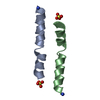
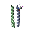
| Deposited unit | 
| ||||||||
|---|---|---|---|---|---|---|---|---|---|
| 1 | 
| ||||||||
| 2 | 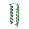
| ||||||||
| 3 |
| ||||||||
| 4 | 
| ||||||||
| Unit cell |
|
-Components
| #1: Protein | Mass: 7241.248 Da / Num. of mol.: 2 / Fragment: Residues 59-116 Source method: isolated from a genetically manipulated source Details: M1-binding domain of NEP / Source: (gene. exp.) Influenza A virus / Genus: Influenzavirus A / Gene: NS2 / Production host:  Escherichia coli (E. coli) / References: UniProt: P03508 Escherichia coli (E. coli) / References: UniProt: P03508 |
|---|
-Experimental details
-Experiment
| Experiment | Method: X-RAY DIFFRACTION |
|---|
- Sample preparation
| Crystal | Density Matthews: 4.62 Å3/Da / Density % sol: 73.2 % | |||||||||||||||
|---|---|---|---|---|---|---|---|---|---|---|---|---|---|---|---|---|
| Crystal grow | Temperature: 298 K / Method: vapor diffusion, hanging drop / pH: 7.5 Details: PROTEIN IN 20 MM TRIS-HCL PH7.5, 150 MM NACL, HANGING DROP METHOD, ROOM TEMPERATURE, vapor diffusion, hanging drop, temperature 298.0K | |||||||||||||||
| Crystal grow | *PLUS | |||||||||||||||
| Components of the solutions | *PLUS
|
-Data collection
| Diffraction | Mean temperature: 100 K |
|---|---|
| Diffraction source | Source: SYNCHROTRON / Site: ESRF  / Beamline: ID14-2 / Wavelength: 0.933 / Beamline: ID14-2 / Wavelength: 0.933 |
| Detector | Type: ADSC QUANTUM 4 / Detector: CCD / Date: Sep 30, 2002 |
| Radiation | Protocol: SINGLE WAVELENGTH / Monochromatic (M) / Laue (L): M / Scattering type: x-ray |
| Radiation wavelength | Wavelength: 0.933 Å / Relative weight: 1 |
| Reflection | Resolution: 2.6→46.6 Å / Num. obs: 7302 / % possible obs: 99.6 % / Redundancy: 3.8 % / Biso Wilson estimate: 48.47 Å2 / Rmerge(I) obs: 0.09 / Rsym value: 0.09 / Net I/σ(I): 6 |
| Reflection shell | Resolution: 2.6→2.74 Å / Redundancy: 3.8 % / Rmerge(I) obs: 0.32 / Mean I/σ(I) obs: 2 / Rsym value: 0.32 / % possible all: 100 |
| Reflection | *PLUS Highest resolution: 2.6 Å / Rmerge(I) obs: 0.09 |
| Reflection shell | *PLUS % possible obs: 100 % |
- Processing
| Software |
| ||||||||||||||||||||||||||||||||||||||||||||||||||||||||||||||||||||||||||||||||||||||||||||||||||||||||||||||||||||||||||||||||||
|---|---|---|---|---|---|---|---|---|---|---|---|---|---|---|---|---|---|---|---|---|---|---|---|---|---|---|---|---|---|---|---|---|---|---|---|---|---|---|---|---|---|---|---|---|---|---|---|---|---|---|---|---|---|---|---|---|---|---|---|---|---|---|---|---|---|---|---|---|---|---|---|---|---|---|---|---|---|---|---|---|---|---|---|---|---|---|---|---|---|---|---|---|---|---|---|---|---|---|---|---|---|---|---|---|---|---|---|---|---|---|---|---|---|---|---|---|---|---|---|---|---|---|---|---|---|---|---|---|---|---|---|
| Refinement | Method to determine structure: MIR / Resolution: 2.6→19.88 Å / Cor.coef. Fo:Fc: 0.952 / Cor.coef. Fo:Fc free: 0.909 / SU B: 7.689 / SU ML: 0.162 / TLS residual ADP flag: LIKELY RESIDUAL / Cross valid method: THROUGHOUT / σ(F): 0 / ESU R: 0.269 / ESU R Free: 0.244
| ||||||||||||||||||||||||||||||||||||||||||||||||||||||||||||||||||||||||||||||||||||||||||||||||||||||||||||||||||||||||||||||||||
| Solvent computation | Ion probe radii: 0.8 Å / Shrinkage radii: 0.8 Å / VDW probe radii: 1.4 Å / Solvent model: BABINET MODEL WITH MASK | ||||||||||||||||||||||||||||||||||||||||||||||||||||||||||||||||||||||||||||||||||||||||||||||||||||||||||||||||||||||||||||||||||
| Displacement parameters | Biso mean: 43.46 Å2
| ||||||||||||||||||||||||||||||||||||||||||||||||||||||||||||||||||||||||||||||||||||||||||||||||||||||||||||||||||||||||||||||||||
| Refinement step | Cycle: LAST / Resolution: 2.6→19.88 Å
| ||||||||||||||||||||||||||||||||||||||||||||||||||||||||||||||||||||||||||||||||||||||||||||||||||||||||||||||||||||||||||||||||||
| Refine LS restraints |
| ||||||||||||||||||||||||||||||||||||||||||||||||||||||||||||||||||||||||||||||||||||||||||||||||||||||||||||||||||||||||||||||||||
| LS refinement shell | Resolution: 2.6→2.67 Å / Total num. of bins used: 20 /
| ||||||||||||||||||||||||||||||||||||||||||||||||||||||||||||||||||||||||||||||||||||||||||||||||||||||||||||||||||||||||||||||||||
| Refinement TLS params. | Method: refined / Refine-ID: X-RAY DIFFRACTION
| ||||||||||||||||||||||||||||||||||||||||||||||||||||||||||||||||||||||||||||||||||||||||||||||||||||||||||||||||||||||||||||||||||
| Refinement TLS group |
| ||||||||||||||||||||||||||||||||||||||||||||||||||||||||||||||||||||||||||||||||||||||||||||||||||||||||||||||||||||||||||||||||||
| Refinement | *PLUS Highest resolution: 2.6 Å / % reflection Rfree: 5 % | ||||||||||||||||||||||||||||||||||||||||||||||||||||||||||||||||||||||||||||||||||||||||||||||||||||||||||||||||||||||||||||||||||
| Solvent computation | *PLUS | ||||||||||||||||||||||||||||||||||||||||||||||||||||||||||||||||||||||||||||||||||||||||||||||||||||||||||||||||||||||||||||||||||
| Displacement parameters | *PLUS | ||||||||||||||||||||||||||||||||||||||||||||||||||||||||||||||||||||||||||||||||||||||||||||||||||||||||||||||||||||||||||||||||||
| Refine LS restraints | *PLUS
|
