 Movie
Movie Controller

[English] 日本語
 Yorodumi
- PDB-1nsk: THE CRYSTAL STRUCTURE OF A HUMAN NUCLEOSIDE DIPHOSPHATE KINASE, N... -
+ Open data

- Basic information
| Entry | Database: PDB / ID: 1nsk | ||||||
|---|---|---|---|---|---|---|---|
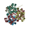
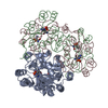
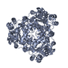
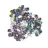
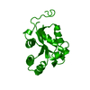
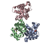
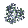
| Title | THE CRYSTAL STRUCTURE OF A HUMAN NUCLEOSIDE DIPHOSPHATE KINASE, NM23-H2 | ||||||
 Components Components | NUCLEOSIDE DIPHOSPHATE KINASE Nucleoside-diphosphate kinase Nucleoside-diphosphate kinase | ||||||
 Keywords Keywords | PHOSPHOTRANSFERASE (PO4 AS ACCEPTOR) | ||||||
| Function / homology |  Function and homology information Function and homology informationregulation of epidermis development / intermediate filament binding / negative regulation of myeloid leukocyte differentiation / nucleoside triphosphate biosynthetic process / protein histidine kinase activity / Ribavirin ADME / G-quadruplex DNA binding / nucleoside-diphosphate kinase / positive regulation of keratinocyte differentiation / Interconversion of nucleotide di- and triphosphates ...regulation of epidermis development / intermediate filament binding / negative regulation of myeloid leukocyte differentiation / nucleoside triphosphate biosynthetic process / protein histidine kinase activity / Ribavirin ADME / G-quadruplex DNA binding / nucleoside-diphosphate kinase / positive regulation of keratinocyte differentiation / Interconversion of nucleotide di- and triphosphates / UTP biosynthetic process / CTP biosynthetic process / Azathioprine ADME / response to growth hormone / GTP biosynthetic process / intermediate filament / nucleoside diphosphate kinase activity / cellular response to fatty acid / histidine kinase / ruffle / positive regulation of epithelial cell proliferation / cell periphery / integrin-mediated signaling pathway / fatty acid binding / cellular response to glucose stimulus / mitochondrial membrane / adenylate cyclase-activating G protein-coupled receptor signaling pathway / positive regulation of neuron projection development / GDP binding / lamellipodium / cellular response to oxidative stress / regulation of apoptotic process / secretory granule lumen / ficolin-1-rich granule lumen / transcription coactivator activity / cell adhesion / protein serine/threonine kinase activity / Neutrophil degranulation / negative regulation of apoptotic process / perinuclear region of cytoplasm / positive regulation of DNA-templated transcription / enzyme binding / positive regulation of transcription by RNA polymerase II / DNA binding / extracellular exosome / extracellular region / ATP binding / identical protein binding / metal ion binding / nucleus / cytosol / cytoplasmSimilarity search - Function | ||||||
| Biological species |  Homo sapiens (human) Homo sapiens (human) | ||||||
| Method | X-RAY DIFFRACTION / Resolution: 2.8 Å | ||||||
 Authors Authors | Williams, R.L. / Perisic, O. | ||||||
 Citation Citation | Journal: J.Mol.Biol. / Year: 1995 Title: The crystal structure of a human nucleoside diphosphate kinase, NM23-H2. Authors: Webb, P.A. / Perisic, O. / Mendola, C.E. / Backer, J.M. / Williams, R.L. | ||||||
| History |
|
- Structure visualization
| Structure viewer | Molecule: MolmilJmol/JSmol |
|---|
- Downloads & links
-Download
| PDBx/mmCIF format | 1nsk.cif.gz | 178.7 KB | Display | PDBx/mmCIF format |
|---|---|---|---|---|
| PDB format | pdb1nsk.ent.gz | 148.2 KB | Display | PDB format |
| PDBx/mmJSON format | 1nsk.json.gz | Tree view | PDBx/mmJSON format | |
| Others |  Other downloads Other downloads |
-Validation report
| Arichive directory | https://data.pdbj.org/pub/pdb/validation_reports/ns/1nskftp://data.pdbj.org/pub/pdb/validation_reports/ns/1nsk | HTTPS FTP |
|---|
-Related structure data
| Similar structure data |
|---|


-Links
 PDBj
PDBj


- Assembly
| Deposited unit | 
| ||||||||||||||||||||||||
|---|---|---|---|---|---|---|---|---|---|---|---|---|---|---|---|---|---|---|---|---|---|---|---|---|---|
| 1 |
| ||||||||||||||||||||||||
| Unit cell |
| ||||||||||||||||||||||||
| Noncrystallographic symmetry (NCS) | NCS oper:
|
-Components
| #1: Protein | Nucleoside-diphosphate kinase Mass: 17324.055 Da / Num. of mol.: 6 Source method: isolated from a genetically manipulated source Source: (gene. exp.) Homo sapiens (human) / Gene: NM23-H2 / Plasmid: PMAL-C2 (NEW ENGLAND BIOLABS) / Gene (production host): NM23-H2 / Production host:  Escherichia coli (E. coli) / References: UniProt: P22392, nucleoside-diphosphate kinase Escherichia coli (E. coli) / References: UniProt: P22392, nucleoside-diphosphate kinase |
|---|
-Experimental details
-Experiment
| Experiment | Method: X-RAY DIFFRACTION / Number of used crystals: 1 |
|---|
- Sample preparation
| Crystal | Density Matthews: 3.06 Å3/Da / Density % sol: 59.76 % | |||||||||||||||||||||||||||||||||||
|---|---|---|---|---|---|---|---|---|---|---|---|---|---|---|---|---|---|---|---|---|---|---|---|---|---|---|---|---|---|---|---|---|---|---|---|---|
| Crystal grow | *PLUS Temperature: 4 ℃ / pH: 8.5 / Method: vapor diffusion, hanging drop | |||||||||||||||||||||||||||||||||||
| Components of the solutions | *PLUS
|
-Data collection
| Diffraction source | Wavelength: 1.5418 Å |
|---|---|
| Detector | Type: MARRESEARCH / Detector: IMAGE PLATE |
| Radiation | Monochromatic (M) / Laue (L): M / Scattering type: x-ray |
| Radiation wavelength | Wavelength: 1.5418 Å / Relative weight: 1 |
| Reflection | Resolution: 2.8→25 Å / Num. obs: 29553 / % possible obs: 92.8 % / Observed criterion σ(I): 0 / Redundancy: 3.4 % / Rmerge(I) obs: 0.087 |
| Reflection | *PLUS Rmerge(I) obs: 0.087 |
- Processing
| Software |
| ||||||||||||||||||||||||||||||||||||||||||||||||||||||||||||
|---|---|---|---|---|---|---|---|---|---|---|---|---|---|---|---|---|---|---|---|---|---|---|---|---|---|---|---|---|---|---|---|---|---|---|---|---|---|---|---|---|---|---|---|---|---|---|---|---|---|---|---|---|---|---|---|---|---|---|---|---|---|
| Refinement | Resolution: 2.8→6 Å / σ(F): 0
| ||||||||||||||||||||||||||||||||||||||||||||||||||||||||||||
| Displacement parameters | Biso mean: 29.63 Å2 | ||||||||||||||||||||||||||||||||||||||||||||||||||||||||||||
| Refinement step | Cycle: LAST / Resolution: 2.8→6 Å
| ||||||||||||||||||||||||||||||||||||||||||||||||||||||||||||
| Refine LS restraints |
|
