 Movie
Movie Controller

[English] 日本語
 Yorodumi
- PDB-1nh4: Structure of the coat protein in fd filamentous bacteriophage par... -
+ Open data

- Basic information
| Entry | Database: PDB / ID: 1nh4 | ||||||
|---|---|---|---|---|---|---|---|
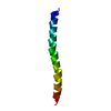
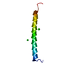
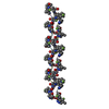
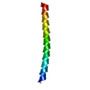
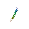
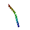
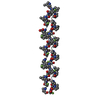
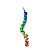
| Title | Structure of the coat protein in fd filamentous bacteriophage particles | ||||||
 Components Components | Major coat protein | ||||||
 Keywords Keywords | VIRUS / ALPHA HELIX / Helical virus | ||||||
| Function / homology |  Function and homology information Function and homology informationhelical viral capsid / host cell membrane / membrane => GO:0016020 / membrane Similarity search - Function | ||||||
| Biological species |  Enterobacteria phage fd (virus) Enterobacteria phage fd (virus) | ||||||
| Method | SOLID-STATE NMR / SOLID-STATE NMR SPECTROSCOPY | ||||||
 Authors Authors | Zeri, A.C. / Mesleh, M.F. / Nevzorov, A.A. / Opella, S.J. | ||||||
 Citation Citation | Journal: Proc.Natl.Acad.Sci.USA / Year: 2003 Title: Structure of the coat protein in fd filamentous bacteriophage particles determined by solid-state NMR spectroscopy Authors: Zeri, A.C. / Mesleh, M.F. / Nevzorov, A.A. / Opella, S.J. | ||||||
| History |
| ||||||
| Remark 999 | Authors informed that the sequence of their protein is identical to residues 27-76 of the database ...Authors informed that the sequence of their protein is identical to residues 27-76 of the database reference sequence provided in GenBank entry 8698956 but that the source of their protein is different. Residue 21 was mutated from Tyr to Met. |
- Structure visualization
| Structure viewer | Molecule: MolmilJmol/JSmol |
|---|
- Downloads & links
-Download
| PDBx/mmCIF format | 1nh4.cif.gz | 264.1 KB | Display | PDBx/mmCIF format |
|---|---|---|---|---|
| PDB format | pdb1nh4.ent.gz | 220 KB | Display | PDB format |
| PDBx/mmJSON format | 1nh4.json.gz | Tree view | PDBx/mmJSON format | |
| Others |  Other downloads Other downloads |
-Validation report
| Summary document | 1nh4_validation.pdf.gz | 333 KB | Display | wwPDB validaton report |
|---|---|---|---|---|
| Full document | 1nh4_full_validation.pdf.gz | 411 KB | Display | |
| Data in XML | 1nh4_validation.xml.gz | 18.3 KB | Display | |
| Data in CIF | 1nh4_validation.cif.gz | 31 KB | Display | |
| Arichive directory | https://data.pdbj.org/pub/pdb/validation_reports/nh/1nh4ftp://data.pdbj.org/pub/pdb/validation_reports/nh/1nh4 | HTTPS FTP |
-Related structure data
| Related structure data | |
|---|---|
| Similar structure data |


-Links
 PDBj
PDBj- Assembly
| Deposited unit | 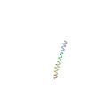
| |||||||||
|---|---|---|---|---|---|---|---|---|---|---|
| 1 |
| |||||||||
| NMR ensembles |
|
-Components
| #1: Protein/peptide | Mass: 5212.021 Da / Num. of mol.: 1 / Mutation: Y21M / Source method: isolated from a natural source / Source: (natural) Enterobacteria phage fd (virus) / Genus: Inovirus / Species: Enterobacteria phage M13 / Strain: fd / References: UniProt: Q9T0Q9, UniProt: P69539*PLUS |
|---|
-Experimental details
-Experiment
| Experiment | Method: SOLID-STATE NMR |
|---|---|
| NMR experiment | Type: PISEMA |
| NMR details | Text: This structure was generated from the five-fold symmetry of PDB entry 1IFI, which has the symmetry that relates one pentamer to the next. This one has had the sidechains added using the program ...Text: This structure was generated from the five-fold symmetry of PDB entry 1IFI, which has the symmetry that relates one pentamer to the next. This one has had the sidechains added using the program SCWRL. The structure was determined by solid state nuclear magnetic resonance using the whole virus in suspension. A model of a section of the virion capsid was built with 20 copies of the protein arranged in agreement with previous studies by fiber diffraction (Marvin et al., J. Mol. Biol. 235, 260286 (1994)). |
- Sample preparation
| Details | Contents: 15N 50mg/ml virus particles in suspension, 5mM sodium borate buffer Solvent system: 100% H2O |
|---|---|
| Sample conditions | pH: 8 / Pressure: ambient / Temperature: 338 K |
| Crystal grow | *PLUS Method: other / Details: NMR |
-NMR measurement
| Radiation | Protocol: SINGLE WAVELENGTH / Monochromatic (M) / Laue (L): M |
|---|---|
| Radiation wavelength | Relative weight: 1 |
| NMR spectrometer | Manufacturer: Home-built / Field strength: 550 MHz |
- Processing
| NMR software |
| ||||||||||||||||
|---|---|---|---|---|---|---|---|---|---|---|---|---|---|---|---|---|---|
| Refinement | Method: SOLID-STATE NMR SPECTROSCOPY / Software ordinal: 1 Details: The first five N-terminal residues of the coat protein undergo isotropic movements at room temperature and structural parameters cannot be resolved. | ||||||||||||||||
| NMR ensemble | Conformers submitted total number: 20 |
