 Movie
Movie Controller

[English] 日本語
 Yorodumi
- PDB-1m5y: Crystallographic Structure of SurA, a Molecular Chaperone that Fa... -
+ Open data

- Basic information
| Entry | Database: PDB / ID: 1m5y | ||||||
|---|---|---|---|---|---|---|---|
| Title | Crystallographic Structure of SurA, a Molecular Chaperone that Facilitates Outer Membrane Porin Folding | ||||||
 Components Components | Survival protein surA | ||||||
 Keywords Keywords | ISOMERASE / CELL CYCLE / Survival protein A / periplasmic molecular chaperone / membrane protein folding / gram negative bacteria | ||||||
| Function / homology |  Function and homology information Function and homology informationmaintenance of stationary phase / maintenance of unfolded protein / Gram-negative-bacterium-type cell outer membrane assembly / chaperone-mediated protein folding / peptidylprolyl isomerase / peptidyl-prolyl cis-trans isomerase activity / peptide binding / unfolded protein binding / protein folding / outer membrane-bounded periplasmic space / protein stabilizationSimilarity search - Function | ||||||
| Biological species |  Escherichia coli (E. coli) Escherichia coli (E. coli) | ||||||
| Method | X-RAY DIFFRACTION / SYNCHROTRON / Resolution: 3 Å | ||||||
 Authors Authors | Bitto, E. / McKay, D.B. | ||||||
 Citation Citation | Journal: Structure / Year: 2002 Title: Crystallographic Structure of SurA, a Molecular Chaperone that Facilitates Folding of Outer Membrane Porins Authors: Bitto, E. / McKay, D.B. | ||||||
| History |
|
- Structure visualization
| Structure viewer | Molecule: MolmilJmol/JSmol |
|---|
- Downloads & links
-Download
| PDBx/mmCIF format | 1m5y.cif.gz | 270.7 KB | Display | PDBx/mmCIF format |
|---|---|---|---|---|
| PDB format | pdb1m5y.ent.gz | 226.6 KB | Display | PDB format |
| PDBx/mmJSON format | 1m5y.json.gz | Tree view | PDBx/mmJSON format | |
| Others |  Other downloads Other downloads |
-Validation report
| Arichive directory | https://data.pdbj.org/pub/pdb/validation_reports/m5/1m5yftp://data.pdbj.org/pub/pdb/validation_reports/m5/1m5y | HTTPS FTP |
|---|
-Related structure data
| Similar structure data |
|---|

-Links
 PDBj
PDBj

- Assembly
| Deposited unit | 
| ||||||||||
|---|---|---|---|---|---|---|---|---|---|---|---|
| 1 | 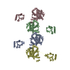
| ||||||||||
| Unit cell |
|
-Components
| #1: Protein | / E.C.5.2.1.8 / survival protein / Peptidyl-prolyl cis-trans isomerase surA / PPIase / Rotamase C Mass: 45131.926 Da / Num. of mol.: 4 Source method: isolated from a genetically manipulated source Source: (gene. exp.) Escherichia coli (E. coli) / Gene: sura / Plasmid: pTYB2 / Species (production host): Escherichia coli / Production host: Escherichia coli BL21(DE3) (bacteria) / Strain (production host): BL21(DE3) / References: UniProt: P0ABZ6, peptidylprolyl isomerase |
|---|
-Experimental details
-Experiment
| Experiment | Method: X-RAY DIFFRACTION / Number of used crystals: 1 |
|---|
- Sample preparation
| Crystal | Density Matthews: 3.43 Å3/Da / Density % sol: 64.19 % | ||||||||||||||||||
|---|---|---|---|---|---|---|---|---|---|---|---|---|---|---|---|---|---|---|---|
| Crystal grow | Temperature: 303 K / Method: vapor diffusion, hanging drop / pH: 5.6 Details: 0.75M ammonium sulphate, 100mM sodium citrate, pH 5.6, VAPOR DIFFUSION, HANGING DROP at 303K, VAPOR DIFFUSION, HANGING DROP, temperature 303.0K | ||||||||||||||||||
| Crystal grow | *PLUS Temperature: 18 ℃ | ||||||||||||||||||
| Components of the solutions | *PLUS
|
-Data collection
| Diffraction |
| ||||||||||||||||||
|---|---|---|---|---|---|---|---|---|---|---|---|---|---|---|---|---|---|---|---|
| Diffraction source |
| ||||||||||||||||||
| Detector |
| ||||||||||||||||||
| Radiation |
| ||||||||||||||||||
| Radiation wavelength | Wavelength: 0.965 Å / Relative weight: 1 | ||||||||||||||||||
| Reflection | Resolution: 3→30 Å / Num. all: 49633 / Num. obs: 45004 / % possible obs: 93.2 % / Observed criterion σ(I): 1 / Redundancy: 4.6 % / Biso Wilson estimate: 59 Å2 / Rsym value: 0.077 / Net I/σ(I): 1.62 | ||||||||||||||||||
| Reflection shell | Resolution: 3→3.05 Å / Mean I/σ(I) obs: 3.9 / Num. unique all: 2114 / Rsym value: 0.348 / % possible all: 85.2 | ||||||||||||||||||
| Reflection | *PLUS Highest resolution: 3 Å / Num. obs: 46785 / % possible obs: 92.9 % / Num. measured all: 217260 / Rmerge(I) obs: 0.077 | ||||||||||||||||||
| Reflection shell | *PLUS % possible obs: 85.2 % / Rmerge(I) obs: 0.348 |

- Processing
| Software |
| ||||||||||||||||||||||||||||||||||||||||||||||||||||||||||||
|---|---|---|---|---|---|---|---|---|---|---|---|---|---|---|---|---|---|---|---|---|---|---|---|---|---|---|---|---|---|---|---|---|---|---|---|---|---|---|---|---|---|---|---|---|---|---|---|---|---|---|---|---|---|---|---|---|---|---|---|---|---|
| Refinement | Resolution: 3→29.8 Å / Rfactor Rfree error: 0.005 / Data cutoff high absF: 1008569.15 / Data cutoff low absF: 0 / Isotropic thermal model: GROUP / Cross valid method: THROUGHOUT / σ(F): 3
| ||||||||||||||||||||||||||||||||||||||||||||||||||||||||||||
| Solvent computation | Solvent model: FLAT MODEL / Bsol: 27.4224 Å2 / ksol: 0.311691 e/Å3 | ||||||||||||||||||||||||||||||||||||||||||||||||||||||||||||
| Displacement parameters | Biso mean: 67.5 Å2
| ||||||||||||||||||||||||||||||||||||||||||||||||||||||||||||
| Refine analyze |
| ||||||||||||||||||||||||||||||||||||||||||||||||||||||||||||
| Refinement step | Cycle: LAST / Resolution: 3→29.8 Å
| ||||||||||||||||||||||||||||||||||||||||||||||||||||||||||||
| Refine LS restraints |
| ||||||||||||||||||||||||||||||||||||||||||||||||||||||||||||
| LS refinement shell | Resolution: 3→3.19 Å / Rfactor Rfree error: 0.018 / Total num. of bins used: 6
| ||||||||||||||||||||||||||||||||||||||||||||||||||||||||||||
| Xplor file | Serial no: 1 / Param file: PROTEIN_REP.PARAM / Topol file: PROTEIN.TOP | ||||||||||||||||||||||||||||||||||||||||||||||||||||||||||||
| Refinement | *PLUS Lowest resolution: 30 Å | ||||||||||||||||||||||||||||||||||||||||||||||||||||||||||||
| Solvent computation | *PLUS | ||||||||||||||||||||||||||||||||||||||||||||||||||||||||||||
| Displacement parameters | *PLUS | ||||||||||||||||||||||||||||||||||||||||||||||||||||||||||||
| Refine LS restraints | *PLUS
| ||||||||||||||||||||||||||||||||||||||||||||||||||||||||||||
| LS refinement shell | *PLUS Lowest resolution: 3.11 Å / Rfactor Rfree: 0.417 / Rfactor Rwork: 0.347 |
