 Movie
Movie Controller

[English] 日本語
 Yorodumi
- PDB-1m0w: Yeast Glutathione Synthase Bound to gamma-glutamyl-cysteine, AMP-... -
+ Open data

- Basic information
| Entry | Database: PDB / ID: 1m0w | ||||||
|---|---|---|---|---|---|---|---|
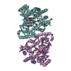
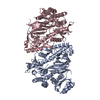
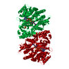
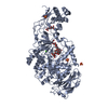
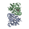
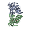
| Title | Yeast Glutathione Synthase Bound to gamma-glutamyl-cysteine, AMP-PNP and 2 Magnesium Ions | ||||||
 Components Components | glutathione synthetase | ||||||
 Keywords Keywords | LIGASE / Amine/Carboxylate Ligase / Structural Genomics / PSI / Protein Structure Initiative / New York SGX Research Center for Structural Genomics / NYSGXRC | ||||||
| Function / homology |  Function and homology information Function and homology informationGlutathione synthesis and recycling / glutathione synthase / glutathione synthase activity / glutathione biosynthetic process / glutathione binding / magnesium ion binding / protein homodimerization activity / ATP binding / cytosol / cytoplasmSimilarity search - Function | ||||||
| Biological species |  Saccharomyces cerevisiae (brewer's yeast) Saccharomyces cerevisiae (brewer's yeast) | ||||||
| Method | X-RAY DIFFRACTION / SYNCHROTRON / MOLECULAR REPLACEMENT / Resolution: 1.8 Å | ||||||
 Authors Authors | Gogos, A. / Shapiro, L. / Burley, S.K. / New York SGX Research Center for Structural Genomics (NYSGXRC) | ||||||
 Citation Citation | Journal: Structure / Year: 2002 Title: Large Conformational Changes in the Catalytic Cycle of Glutathione Synthase Authors: Gogos, A. / Shapiro, L. | ||||||
| History |
|
- Structure visualization
| Structure viewer | Molecule: MolmilJmol/JSmol |
|---|
- Downloads & links
-Download
| PDBx/mmCIF format | 1m0w.cif.gz | 231.4 KB | Display | PDBx/mmCIF format |
|---|---|---|---|---|
| PDB format | pdb1m0w.ent.gz | 180.9 KB | Display | PDB format |
| PDBx/mmJSON format | 1m0w.json.gz | Tree view | PDBx/mmJSON format | |
| Others |  Other downloads Other downloads |
-Validation report
| Arichive directory | https://data.pdbj.org/pub/pdb/validation_reports/m0/1m0wftp://data.pdbj.org/pub/pdb/validation_reports/m0/1m0w | HTTPS FTP |
|---|
-Related structure data
| Related structure data |  1m0tSC S: Starting model for refinement C: citing same article ( |
|---|---|
| Similar structure data | |
| Other databases |


-Links
 PDBj
PDBj


- Assembly
| Deposited unit | 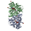
| ||||||||
|---|---|---|---|---|---|---|---|---|---|
| 1 |
| ||||||||
| Unit cell |
|
-Components
-Protein , 1 types, 2 molecules AB
| #1: Protein | / GLUTATHIONE SYNTHASE / GSH SYNTHETASE / GSH-S Mass: 55878.941 Da / Num. of mol.: 2 Source method: isolated from a genetically manipulated source Source: (gene. exp.) Saccharomyces cerevisiae (brewer's yeast)Gene: GSH2 / Plasmid: pET28TEV / Species (production host): Escherichia coli / Production host:  Escherichia coli BL21 (bacteria) / Strain (production host): BL21 / References: UniProt: Q08220, glutathione synthase Escherichia coli BL21 (bacteria) / Strain (production host): BL21 / References: UniProt: Q08220, glutathione synthase |
|---|
-Non-polymers , 5 types, 762 molecules 


| #2: Chemical | ChemComp-MG /  Mass: 24.305 Da / Num. of mol.: 4 / Source method: obtained synthetically / Formula: Mg Mass: 24.305 Da / Num. of mol.: 4 / Source method: obtained synthetically / Formula: Mg#3: Chemical | ChemComp-SO4 / Sulfate Mass: 96.063 Da / Num. of mol.: 7 / Source method: obtained synthetically / Formula: SO4 Mass: 96.063 Da / Num. of mol.: 7 / Source method: obtained synthetically / Formula: SO4#4: Chemical |  Mass: 506.196 Da / Num. of mol.: 2 / Source method: obtained synthetically / Formula: C10H17N6O12P3 / Comment: AMP-PNP, energy-carrying molecule analogue*YM Mass: 506.196 Da / Num. of mol.: 2 / Source method: obtained synthetically / Formula: C10H17N6O12P3 / Comment: AMP-PNP, energy-carrying molecule analogue*YM#5: Chemical | Gamma-L-Glutamyl-L-cysteine Mass: 250.272 Da / Num. of mol.: 2 / Source method: obtained synthetically / Formula: C8H14N2O5S Mass: 250.272 Da / Num. of mol.: 2 / Source method: obtained synthetically / Formula: C8H14N2O5S#6: Water | ChemComp-HOH / | WaterMass: 18.015 Da / Num. of mol.: 747 / Source method: isolated from a natural source / Formula: H2O |
|---|
-Experimental details
-Experiment
| Experiment | Method: X-RAY DIFFRACTION / Number of used crystals: 1 |
|---|
- Sample preparation
| Crystal | Density Matthews: 2.34 Å3/Da / Density % sol: 47.38 % | |||||||||||||||||||||||||||||||||||||||||||||||||||||||||||||||
|---|---|---|---|---|---|---|---|---|---|---|---|---|---|---|---|---|---|---|---|---|---|---|---|---|---|---|---|---|---|---|---|---|---|---|---|---|---|---|---|---|---|---|---|---|---|---|---|---|---|---|---|---|---|---|---|---|---|---|---|---|---|---|---|---|
| Crystal grow | Temperature: 295 K / Method: vapor diffusion, hanging drop / pH: 8 Details: Ammonium sulfate, PEG 400, Tris-HCl pH 8.0, VAPOR DIFFUSION, HANGING DROP, temperature 295K | |||||||||||||||||||||||||||||||||||||||||||||||||||||||||||||||
| Crystal grow | *PLUS Temperature: 22 ℃ | |||||||||||||||||||||||||||||||||||||||||||||||||||||||||||||||
| Components of the solutions | *PLUS
|
-Data collection
| Diffraction | Mean temperature: 100 K |
|---|---|
| Diffraction source | Source: SYNCHROTRON / Site: NSLS  / Beamline: X4A / Wavelength: 1.04 Å / Beamline: X4A / Wavelength: 1.04 Å |
| Detector | Type: ADSC QUANTUM 4 / Detector: CCD / Date: Oct 23, 2001 / Details: double crystal monochromator with sagital focusing |
| Radiation | Monochromator: Si / Protocol: SINGLE WAVELENGTH / Monochromatic (M) / Laue (L): M / Scattering type: x-ray |
| Radiation wavelength | Wavelength: 1.04 Å / Relative weight: 1 |
| Reflection | Resolution: 1.8→20 Å / Num. all: 91335 / Num. obs: 91335 / % possible obs: 97.2 % / Observed criterion σ(F): 0 / Observed criterion σ(I): -3 / Redundancy: 2.8 % / Biso Wilson estimate: 15.9 Å2 / Rmerge(I) obs: 0.066 / Rsym value: 0.066 / Net I/σ(I): 15.17 |
| Reflection shell | Resolution: 1.8→1.86 Å / Redundancy: 2.8 % / Rmerge(I) obs: 0.4 / Mean I/σ(I) obs: 4.4 / Num. unique all: 8973 / Rsym value: 0.4 / % possible all: 95.4 |
| Reflection | *PLUS Lowest resolution: 20 Å / Num. measured all: 505425 |
| Reflection shell | *PLUS % possible obs: 95.4 % / Rmerge(I) obs: 0.4 |
- Processing
| Software |
| |||||||||||||||||||||||||
|---|---|---|---|---|---|---|---|---|---|---|---|---|---|---|---|---|---|---|---|---|---|---|---|---|---|---|
| Refinement | Method to determine structure: MOLECULAR REPLACEMENT Starting model: 1M0T Resolution: 1.8→20 Å / Rfactor Rfree error: 0.003 / Isotropic thermal model: RESTRAINED / Cross valid method: THROUGHOUT / σ(F): 0 / σ(I): 0 / Stereochemistry target values: Engh & Huber
| |||||||||||||||||||||||||
| Solvent computation | Solvent model: FLAT MODEL / Bsol: 68.0262 Å2 / ksol: 0.390124 e/Å3 | |||||||||||||||||||||||||
| Displacement parameters | Biso mean: 20.8 Å2
| |||||||||||||||||||||||||
| Refine analyze |
| |||||||||||||||||||||||||
| Refinement step | Cycle: LAST / Resolution: 1.8→20 Å
| |||||||||||||||||||||||||
| Refine LS restraints |
| |||||||||||||||||||||||||
| LS refinement shell | Resolution: 1.8→1.91 Å / Rfactor Rfree error: 0.01 / Total num. of bins used: 6
| |||||||||||||||||||||||||
| Xplor file |
| |||||||||||||||||||||||||
| Refinement | *PLUS Highest resolution: 1.82 Å / Lowest resolution: 20 Å / % reflection Rfree: 5 % | |||||||||||||||||||||||||
| Solvent computation | *PLUS | |||||||||||||||||||||||||
| Displacement parameters | *PLUS | |||||||||||||||||||||||||
| Refine LS restraints | *PLUS
|
