 Movie
Movie Controller

[English] 日本語
 Yorodumi
- PDB-1lma: PROTEIN HYDRATION AND WATER STRUCTURE: X-RAY ANALYSIS OF A CLOSEL... -
+ Open data

- Basic information
| Entry | Database: PDB / ID: 1lma | ||||||
|---|---|---|---|---|---|---|---|
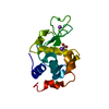
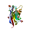
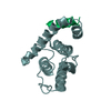
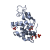
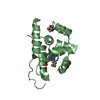
| Title | PROTEIN HYDRATION AND WATER STRUCTURE: X-RAY ANALYSIS OF A CLOSELY PACKED PROTEIN CRYSTAL WITH VERY LOW SOLVENT CONTENT | ||||||
 Components Components | HEN EGG WHITE LYSOZYME | ||||||
 Keywords Keywords | HYDROLASE(O-GLYCOSYL) | ||||||
| Function / homology |  Function and homology information Function and homology informationLactose synthesis / Antimicrobial peptides / Neutrophil degranulation / beta-N-acetylglucosaminidase activity / cell wall macromolecule catabolic process / lysozyme / lysozyme activity / defense response to Gram-negative bacterium / killing of cells of another organism / defense response to Gram-positive bacterium ...Lactose synthesis / Antimicrobial peptides / Neutrophil degranulation / beta-N-acetylglucosaminidase activity / cell wall macromolecule catabolic process / lysozyme / lysozyme activity / defense response to Gram-negative bacterium / killing of cells of another organism / defense response to Gram-positive bacterium / defense response to bacterium / endoplasmic reticulum / extracellular space / identical protein binding / cytoplasm Similarity search - Function | ||||||
| Biological species |  | ||||||
| Method |  X-RAY DIFFRACTION / Resolution: 1.75 Å X-RAY DIFFRACTION / Resolution: 1.75 Å | ||||||
 Authors Authors | Madhusudan / Kodandapani, R. / Vijayan, M. | ||||||
 Citation Citation | Journal: Acta Crystallogr.,Sect.D / Year: 1993 Title: Protein hydration and water structure: X-ray analysis of a closely packed protein crystal with very low solvent content. Authors: Madhusudan / Kodandapani, R. / Vijayan, M. #1: Journal: Curr.Sci. / Year: 1991Title: Rigid and Flexible Regions in Lysozyme and the Invariant Features in its Hydration Shell Authors: Madhusudan / Vijayan, M. #2: Journal: J.Biol.Chem. / Year: 1990Title: Crystal Structure of Low Humidity Tetragonal Lysozyme at 2.1 Angstroms Resolution: Variability in Hydration Shell and its Structural Consequences Authors: Kodandapani, R. / Suresh, C.G. / Vijayan, M. #3: Journal: Acta Crystallogr.,Sect.B / Year: 1985Title: Water-Mediated Transformations in Protein Crystals Authors: Salunke, D.M. / Veerapandian, B. / Kodandapani, R. / Vijayan, M. #4: Journal: Curr.Sci. / Year: 1984Title: Water-Mediated Structural Transformations in a New Crystal Form of Ribonuclease A and Tetragonal Lysozyme Authors: Salunke, D.M. / Veerapandian, B. / Vijayan, M. | ||||||
| History |
|
- Structure visualization
| Structure viewer | Molecule: MolmilJmol/JSmol |
|---|
- Downloads & links
-Download
| PDBx/mmCIF format | 1lma.cif.gz | 35.9 KB | Display | PDBx/mmCIF format |
|---|---|---|---|---|
| PDB format | pdb1lma.ent.gz | 27.9 KB | Display | PDB format |
| PDBx/mmJSON format | 1lma.json.gz | Tree view | PDBx/mmJSON format | |
| Others |  Other downloads Other downloads |
-Validation report
| Summary document | 1lma_validation.pdf.gz | 433.8 KB | Display | wwPDB validaton report |
|---|---|---|---|---|
| Full document | 1lma_full_validation.pdf.gz | 437.3 KB | Display | |
| Data in XML | 1lma_validation.xml.gz | 9.7 KB | Display | |
| Data in CIF | 1lma_validation.cif.gz | 13.1 KB | Display | |
| Arichive directory | https://data.pdbj.org/pub/pdb/validation_reports/lm/1lmaftp://data.pdbj.org/pub/pdb/validation_reports/lm/1lma | HTTPS FTP |
-Related structure data
| Similar structure data |
|---|


-Links
 PDBj
PDBj


- Assembly
| Deposited unit | 
| ||||||||
|---|---|---|---|---|---|---|---|---|---|
| 1 |
| ||||||||
| Unit cell |
| ||||||||
| Atom site foot note | 1: THE ELECTRON DENSITY OF SIDE CHAIN ATOMS OF GLN 121 IS TOO POOR TO ACCURATELY POSITION SIDE CHAIN ATOMS BEYOND CB. |
-Components
| #1: Protein | Mass: 14331.160 Da / Num. of mol.: 1 Source method: isolated from a genetically manipulated source Source: (gene. exp.) | ||
|---|---|---|---|
| #2: Chemical |   Mass: 62.005 Da / Num. of mol.: 2 / Source method: obtained synthetically / Formula: NO3 Mass: 62.005 Da / Num. of mol.: 2 / Source method: obtained synthetically / Formula: NO3#3: Water | ChemComp-HOH / |  Mass: 18.015 Da / Num. of mol.: 148 / Source method: isolated from a natural source / Formula: H2O Mass: 18.015 Da / Num. of mol.: 148 / Source method: isolated from a natural source / Formula: H2O |
-Experimental details
-Experiment
| Experiment | Method: X-RAY DIFFRACTION |
|---|
- Sample preparation
| Crystal | Density Matthews: 1.61 Å3/Da / Density % sol: 23.5 % | |||||||||||||||
|---|---|---|---|---|---|---|---|---|---|---|---|---|---|---|---|---|
| Crystal grow | *PLUS pH: 4.5 / Method: unknownDetails: taken from Steinrauf, L.K. (1959). Acta Cryst. 12, 77. | |||||||||||||||
| Components of the solutions | *PLUS
|
-Data collection
| Radiation | Scattering type: x-ray |
|---|---|
| Radiation wavelength | Relative weight: 1 |
| Reflection | *PLUS Highest resolution: 1.75 Å / Num. obs: 8288 / Observed criterion σ(I): 2 / Num. measured all: 43889 / Rmerge(I) obs: 0.061 / Biso Wilson estimate: 10 Å2 |
- Processing
| Software | Name: PROLSQ / Classification: refinement | ||||||||||||||||||||||||||||||||||||||||||||||||||||||||||||||||||||||||||||||||||||
|---|---|---|---|---|---|---|---|---|---|---|---|---|---|---|---|---|---|---|---|---|---|---|---|---|---|---|---|---|---|---|---|---|---|---|---|---|---|---|---|---|---|---|---|---|---|---|---|---|---|---|---|---|---|---|---|---|---|---|---|---|---|---|---|---|---|---|---|---|---|---|---|---|---|---|---|---|---|---|---|---|---|---|---|---|---|
| Refinement | Resolution: 1.75→10 Å / σ(F): 4 /
| ||||||||||||||||||||||||||||||||||||||||||||||||||||||||||||||||||||||||||||||||||||
| Refinement step | Cycle: LAST / Resolution: 1.75→10 Å
| ||||||||||||||||||||||||||||||||||||||||||||||||||||||||||||||||||||||||||||||||||||
| Refine LS restraints |
| ||||||||||||||||||||||||||||||||||||||||||||||||||||||||||||||||||||||||||||||||||||
| Software | *PLUS Name: PROLSQ / Classification: refinement | ||||||||||||||||||||||||||||||||||||||||||||||||||||||||||||||||||||||||||||||||||||
| Refinement | *PLUS Highest resolution: 1.75 Å / Lowest resolution: 10 Å / Num. reflection obs: 7684 / σ(F): 4 / Rfactor obs: 0.175 | ||||||||||||||||||||||||||||||||||||||||||||||||||||||||||||||||||||||||||||||||||||
| Solvent computation | *PLUS | ||||||||||||||||||||||||||||||||||||||||||||||||||||||||||||||||||||||||||||||||||||
| Displacement parameters | *PLUS |
