 Movie
Movie Controller

[English] 日本語
 Yorodumi
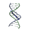
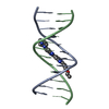
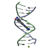
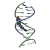
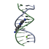
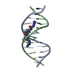
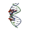
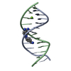
- PDB-1ley: STRUCTURE OF A DICATIONIC MONOIMIDAZOLE LEXITROPSIN BOUND TO DNA ... -
+ Open data

- Basic information
| Entry | Database: PDB / ID: 1ley | ||||||||||||||||||
|---|---|---|---|---|---|---|---|---|---|---|---|---|---|---|---|---|---|---|---|
| Title | STRUCTURE OF A DICATIONIC MONOIMIDAZOLE LEXITROPSIN BOUND TO DNA (ORIENTATION 2) | ||||||||||||||||||
 Components Components | DNA (5'-D(* Keywords Keywords DNA / B-DNA / DOUBLE HELIX / COMPLEXED WITH DRUG DNA / B-DNA / DOUBLE HELIX / COMPLEXED WITH DRUGFunction / homology | MONOIMIDAZOLE LEXITROPSIN / | DNA / DNA (> 10) Function and homology information Function and homology informationMethod | X-RAY DIFFRACTION / Resolution: 2.25 Å  Authors AuthorsGoodsell, D.S. / Ng, H.L. / Kopka, M.L. / Lown, J.W. / Dickerson, R.E. |  CitationJournal: Biochemistry / Year: 1995 CitationJournal: Biochemistry / Year: 1995Title: Structure of a dicationic monoimidazole lexitropsin bound to DNA. Authors: Goodsell, D.S. / Ng, H.L. / Kopka, M.L. / Lown, J.W. / Dickerson, R.E. History |
|
- Structure visualization
| Structure viewer | Molecule: MolmilJmol/JSmol |
|---|
- Downloads & links
-Download
| PDBx/mmCIF format | 1ley.cif.gz | 26.8 KB | Display | PDBx/mmCIF format |
|---|---|---|---|---|
| PDB format | pdb1ley.ent.gz | 16 KB | Display | PDB format |
| PDBx/mmJSON format | 1ley.json.gz | Tree view | PDBx/mmJSON format | |
| Others |  Other downloads Other downloads |
-Validation report
| Arichive directory | https://data.pdbj.org/pub/pdb/validation_reports/le/1leyftp://data.pdbj.org/pub/pdb/validation_reports/le/1ley | HTTPS FTP |
|---|
-Related structure data


-Links
 PDBj
PDBj


- Assembly
| Deposited unit | 
| ||||||||
|---|---|---|---|---|---|---|---|---|---|
| 1 |
| ||||||||
| Unit cell |
| ||||||||
| Details | THIS ENTRY CONTAINS THE CRYSTALLOGRAPHIC ASYMMETRIC UNIT WHICH CONSISTS OF 2 DNA CHAINS AND 1 MONOIMIDAZOLE LEXITROPSIN MOLECULE. INDEPENDENT REFINEMENTS OF THE STRUCTURE WERE CARRIED OUT WITH THE LEXITROPSIN MOLECULE IN TWO DIFFERENT ORIENTATIONS. IT COULD NOT BE DETERMINED WHICH OF THESE ORIENTATIONS WAS CORRECT. THE TWO SETS OF COORDINATES ARE AVAILABLE IN THE PDB FILES 1LEX AND 1LEY. |
-Components
| #1: DNA chain | Mass: 3663.392 Da / Num. of mol.: 2 / Source method: obtained synthetically #2: Chemical | ChemComp-ILT / |   Mass: 431.452 Da / Num. of mol.: 1 / Source method: obtained synthetically / Formula: C17H25N11O3 Mass: 431.452 Da / Num. of mol.: 1 / Source method: obtained synthetically / Formula: C17H25N11O3#3: Water | ChemComp-HOH / | Water Mass: 18.015 Da / Num. of mol.: 37 / Source method: isolated from a natural source / Formula: H2O Mass: 18.015 Da / Num. of mol.: 37 / Source method: isolated from a natural source / Formula: H2O |
|---|
-Experimental details
-Experiment
| Experiment | Method: X-RAY DIFFRACTION |
|---|
- Sample preparation
| Crystal | Density Matthews: 2.04 Å3/Da / Density % sol: 39.85 % | |||||||||||||||||||||||||||||||||||
|---|---|---|---|---|---|---|---|---|---|---|---|---|---|---|---|---|---|---|---|---|---|---|---|---|---|---|---|---|---|---|---|---|---|---|---|---|
| Crystal grow | Temperature: 278 K / Method: vapor diffusion, sitting drop / Details: VAPOR DIFFUSION, SITTING DROP, temperature 278.00K | |||||||||||||||||||||||||||||||||||
| Components of the solutions |
| |||||||||||||||||||||||||||||||||||
| Crystal grow | *PLUS Temperature: 5 ℃ / Method: vapor diffusion | |||||||||||||||||||||||||||||||||||
| Components of the solutions | *PLUS
|
-Data collection
| Diffraction | Mean temperature: 248 K |
|---|---|
| Detector | Type: RIGAKU AFC-5R / Detector: DIFFRACTOMETER |
| Radiation | Protocol: SINGLE WAVELENGTH / Monochromatic (M) / Laue (L): M / Scattering type: x-ray |
| Radiation wavelength | Relative weight: 1 |
| Reflection | Highest resolution: 2.25 Å |
- Processing
| Software | Name: NUCLSQ / Classification: refinement | ||||||||||||||||||||||||||||||||||||||||||||||||||||||||||||||||||||||||||||||||||||
|---|---|---|---|---|---|---|---|---|---|---|---|---|---|---|---|---|---|---|---|---|---|---|---|---|---|---|---|---|---|---|---|---|---|---|---|---|---|---|---|---|---|---|---|---|---|---|---|---|---|---|---|---|---|---|---|---|---|---|---|---|---|---|---|---|---|---|---|---|---|---|---|---|---|---|---|---|---|---|---|---|---|---|---|---|---|
| Refinement | Resolution: 2.25→8 Å / σ(F): 2
| ||||||||||||||||||||||||||||||||||||||||||||||||||||||||||||||||||||||||||||||||||||
| Refinement step | Cycle: LAST / Resolution: 2.25→8 Å
| ||||||||||||||||||||||||||||||||||||||||||||||||||||||||||||||||||||||||||||||||||||
| Refine LS restraints |
|
