 ムービー
ムービー コントローラー


+ データを開く

- 基本情報
基本情報
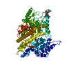
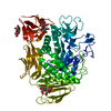
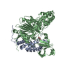
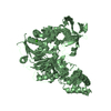
| 登録情報 | データベース: PDB / ID: 1keh | ||||||
|---|---|---|---|---|---|---|---|
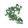
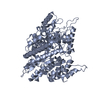
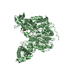
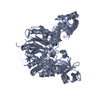
| タイトル | Precursor structure of cephalosporin acylase | ||||||
 要素 要素 | precursor of cephalosporin acylase | ||||||
 キーワード キーワード | HYDROLASE / cephalosporin acylase / precursor / glutaryl-7-ACA | ||||||
| 機能・相同性 |  機能・相同性情報 機能・相同性情報glutaryl-7-aminocephalosporanic-acid acylase / glutaryl-7-aminocephalosporanic-acid acylase activity / antibiotic biosynthetic process / periplasmic space / response to antibiotic 類似検索 - 分子機能 | ||||||
| 生物種 |  Brevundimonas diminuta (バクテリア) Brevundimonas diminuta (バクテリア) | ||||||
| 手法 |  X線回折 / シンクロトロン / フーリエ合成 / 解像度: 2.5 Å X線回折 / シンクロトロン / フーリエ合成 / 解像度: 2.5 Å | ||||||
 データ登録者 データ登録者 | Kim, Y. / Kim, S. | ||||||
 引用 引用 | ジャーナル: J.Biol.Chem. / 年: 2002 タイトル: Precursor structure of cephalosporin acylase. Insights into autoproteolytic activation in a new N-terminal hydrolase family 著者: Kim, Y. / Kim, S. / Earnest, T.N. / Hol, W.G. | ||||||
| 履歴 |
|
- 構造の表示
構造の表示
| 構造ビューア | 分子: MolmilJmol/JSmol |
|---|
- ダウンロードとリンク
-ダウンロード
| PDBx/mmCIF形式 | 1keh.cif.gz | 151.3 KB | 表示 | PDBx/mmCIF形式 |
|---|---|---|---|---|
| PDB形式 | pdb1keh.ent.gz | 118.4 KB | 表示 | PDB形式 |
| PDBx/mmJSON形式 | 1keh.json.gz | ツリー表示 | PDBx/mmJSON形式 | |
| その他 |  その他のダウンロード その他のダウンロード |
-検証レポート
| アーカイブディレクトリ | https://data.pdbj.org/pub/pdb/validation_reports/ke/1kehftp://data.pdbj.org/pub/pdb/validation_reports/ke/1keh | HTTPS FTP |
|---|
-関連構造データ
-リンク
 PDBj
PDBj


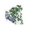
- 集合体
集合体
| 登録構造単位 | 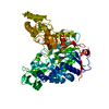
| ||||||||
|---|---|---|---|---|---|---|---|---|---|
| 1 |
| ||||||||
| 単位格子 |
|
-要素
| #1: タンパク質 | 分子量: 76609.883 Da / 分子数: 1 / 断片: residues 1-689 / 変異: S170A,R428A / 由来タイプ: 組換発現 由来: (組換発現) Brevundimonas diminuta (バクテリア)プラスミド: pET24d(+) / 発現宿主: 参照: UniProt: Q9L5D6, 加水分解酵素; ペプチド以外のCN結合加水分解酵素; 鎖状アミドに作用 |
|---|---|
| #2: 水 | ChemComp-HOH /  分子量: 18.015 Da / 分子数: 399 / 由来タイプ: 天然 / 式: H2O 分子量: 18.015 Da / 分子数: 399 / 由来タイプ: 天然 / 式: H2O |
-実験情報
-実験
| 実験 | 手法: X線回折 / 使用した結晶の数: 1 |
|---|
- 試料調製
試料調製
| 結晶 | マシュー密度: 3.37 Å3/Da / 溶媒含有率: 63.55 % | ||||||||||||||||||||||||||||||||||||||||||||||||||||||||
|---|---|---|---|---|---|---|---|---|---|---|---|---|---|---|---|---|---|---|---|---|---|---|---|---|---|---|---|---|---|---|---|---|---|---|---|---|---|---|---|---|---|---|---|---|---|---|---|---|---|---|---|---|---|---|---|---|---|
| 結晶化 | 温度: 290 K / 手法: 蒸気拡散法, ハンギングドロップ法 / pH: 6.5 詳細: PEG8000, MgAcetate, Sodium Cacodylate, DTT, pH 6.5, VAPOR DIFFUSION, HANGING DROP, temperature 290K | ||||||||||||||||||||||||||||||||||||||||||||||||||||||||
| 結晶化 | *PLUS 温度: 21 ℃ / pH: 7 | ||||||||||||||||||||||||||||||||||||||||||||||||||||||||
| 溶液の組成 | *PLUS
|
-データ収集
| 回折 | 平均測定温度: 125 K |
|---|---|
| 放射光源 | 由来: シンクロトロン / サイト: ALS  / ビームライン: 5.0.2 / 波長: 0.91 Å / ビームライン: 5.0.2 / 波長: 0.91 Å |
| 検出器 | タイプ: CUSTOM-MADE / 検出器: CCD / 日付: 2001年1月7日 |
| 放射 | モノクロメーター: graphite / プロトコル: SINGLE WAVELENGTH / 単色(M)・ラウエ(L): M / 散乱光タイプ: x-ray |
| 放射波長 | 波長: 0.91 Å / 相対比: 1 |
| 反射 | 解像度: 2.5→20 Å / Num. all: 39792 / Num. obs: 38001 / % possible obs: 95.5 % / Observed criterion σ(F): 0 / Observed criterion σ(I): 3.2 |
| 反射 シェル | 解像度: 2.5→2.59 Å / % possible all: 70.4 |
| 反射 | *PLUS 最低解像度: 20 Å / Num. measured all: 838095 / Rmerge(I) obs: 0.108 |
| 反射 シェル | *PLUS 最高解像度: 2.5 Å / % possible obs: 70.4 % / Rmerge(I) obs: 0.548 / Mean I/σ(I) obs: 3.2 |
- 解析
解析
| ソフトウェア |
| |||||||||||||||||||||||||
|---|---|---|---|---|---|---|---|---|---|---|---|---|---|---|---|---|---|---|---|---|---|---|---|---|---|---|
| 精密化 | 構造決定の手法: フーリエ合成 / 解像度: 2.5→20 Å / σ(F): 0 / 立体化学のターゲット値: Engh & Huber
| |||||||||||||||||||||||||
| 精密化ステップ | サイクル: LAST / 解像度: 2.5→20 Å
| |||||||||||||||||||||||||
| 精密化 | *PLUS 最低解像度: 20 Å / % reflection Rfree: 3.5 % / Rfactor obs: 0.203 | |||||||||||||||||||||||||
| 溶媒の処理 | *PLUS | |||||||||||||||||||||||||
| 原子変位パラメータ | *PLUS | |||||||||||||||||||||||||
| 拘束条件 | *PLUS
|
