 Movie
Movie Controller

[English] 日本語
 Yorodumi
- PDB-1k4o: Crystal Structure of 3,4-dihydroxy-2-butanone 4-phosphate synthas... -
+ Open data

- Basic information
| Entry | Database: PDB / ID: 1k4o | ||||||
|---|---|---|---|---|---|---|---|
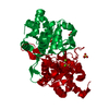
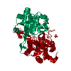
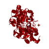
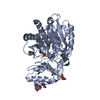
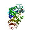
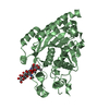
| Title | Crystal Structure of 3,4-dihydroxy-2-butanone 4-phosphate synthase in complex with one Manganese, and a glycerol | ||||||
 Components Components | 3,4-Dihydroxy-2-Butanone 4-Phosphate Synthase | ||||||
 Keywords Keywords | ISOMERASE / dihydroxybutanone phosphate synthase / riboflavin biosynthesis / antimicrobial target / structure-based design | ||||||
| Function / homology |  Function and homology information Function and homology information3,4-dihydroxy-2-butanone-4-phosphate synthase / 3,4-dihydroxy-2-butanone-4-phosphate synthase activity / riboflavin biosynthetic process / metal ion binding / cytosol Similarity search - Function | ||||||
| Biological species |  Magnaporthe grisea (fungus) Magnaporthe grisea (fungus) | ||||||
| Method |  X-RAY DIFFRACTION / SYNCHROTRON / FOURIER SYNTHESIS / Resolution: 1.1 Å X-RAY DIFFRACTION / SYNCHROTRON / FOURIER SYNTHESIS / Resolution: 1.1 Å | ||||||
 Authors Authors | Liao, D.-I. / Zheng, Y.-J. / Viitanen, P.V. / Jordan, D.B. | ||||||
 Citation Citation | Journal: Biochemistry / Year: 2002 Title: Structural definition of the active site and catalytic mechanism of 3,4-dihydroxy-2-butanone-4-phosphate synthase. Authors: Liao, D.I. / Zheng, Y.J. / Viitanen, P.V. / Jordan, D.B. | ||||||
| History |
|
- Structure visualization
| Structure viewer | Molecule: MolmilJmol/JSmol |
|---|
- Downloads & links
-Download
| PDBx/mmCIF format | 1k4o.cif.gz | 61.7 KB | Display | PDBx/mmCIF format |
|---|---|---|---|---|
| PDB format | pdb1k4o.ent.gz | 42.7 KB | Display | PDB format |
| PDBx/mmJSON format | 1k4o.json.gz | Tree view | PDBx/mmJSON format | |
| Others |  Other downloads Other downloads |
-Validation report
| Summary document | 1k4o_validation.pdf.gz | 390.2 KB | Display | wwPDB validaton report |
|---|---|---|---|---|
| Full document | 1k4o_full_validation.pdf.gz | 395.2 KB | Display | |
| Data in XML | 1k4o_validation.xml.gz | 6.4 KB | Display | |
| Data in CIF | 1k4o_validation.cif.gz | 10.5 KB | Display | |
| Arichive directory | https://data.pdbj.org/pub/pdb/validation_reports/k4/1k4oftp://data.pdbj.org/pub/pdb/validation_reports/k4/1k4o | HTTPS FTP |
-Related structure data
| Related structure data |  1k49SC  1k4iC  1k4lC  1k4pC S: Starting model for refinement C: citing same article ( |
|---|---|
| Similar structure data |
-Links
 PDBj
PDBj- Assembly
| Deposited unit | 
| ||||||||
|---|---|---|---|---|---|---|---|---|---|
| 1 | 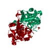
| ||||||||
| Unit cell |
| ||||||||
| Components on special symmetry positions |
| ||||||||
| Details | The biological assembly is a homodimer. Applying the following symmetry related operation on the coordinates of the monomer in the asymmetric unit would generate the 2nd half of the dimer. rotation matrix -1.00000 0.00000 0.00000 0.00000 -1.00000 0.00000 0.00000 0.00000 1.00000 translation -1, -1, 0 |
-Components
| #1: Protein | Mass: 25036.338 Da / Num. of mol.: 1 Source method: isolated from a genetically manipulated source Source: (gene. exp.) Magnaporthe grisea (fungus) / Gene: rice blast fungi / Plasmid: PET-24a(+) / Species (production host): Escherichia coli / Production host:  References: UniProt: Q8TG90, Isomerases; Intramolecular transferases; Transferring other groups | ||||||
|---|---|---|---|---|---|---|---|
| #2: Chemical |   Mass: 96.063 Da / Num. of mol.: 2 / Source method: obtained synthetically / Formula: SO4 Mass: 96.063 Da / Num. of mol.: 2 / Source method: obtained synthetically / Formula: SO4#3: Chemical | ChemComp-MN / |   Mass: 54.938 Da / Num. of mol.: 1 / Source method: obtained synthetically / Formula: Mn Mass: 54.938 Da / Num. of mol.: 1 / Source method: obtained synthetically / Formula: Mn#4: Chemical | ChemComp-GOL / |   Mass: 92.094 Da / Num. of mol.: 1 / Source method: obtained synthetically / Formula: C3H8O3 Mass: 92.094 Da / Num. of mol.: 1 / Source method: obtained synthetically / Formula: C3H8O3#5: Water | ChemComp-HOH / |  Mass: 18.015 Da / Num. of mol.: 237 / Source method: isolated from a natural source / Formula: H2O Mass: 18.015 Da / Num. of mol.: 237 / Source method: isolated from a natural source / Formula: H2O |
-Experimental details
-Experiment
| Experiment | Method: X-RAY DIFFRACTION / Number of used crystals: 1 |
|---|
- Sample preparation
| Crystal | Density Matthews: 2.07 Å3/Da / Density % sol: 40.47 % | |||||||||||||||||||||||||||||||||||
|---|---|---|---|---|---|---|---|---|---|---|---|---|---|---|---|---|---|---|---|---|---|---|---|---|---|---|---|---|---|---|---|---|---|---|---|---|
| Crystal grow | Temperature: 295 K / Method: vapor diffusion, hanging drop / pH: 6.5 Details: Li2SO4, MES-NaOH, PEG5000 Monomethyl ether, MnCl2, pH 6.5, VAPOR DIFFUSION, HANGING DROP, temperature 295K | |||||||||||||||||||||||||||||||||||
| Crystal grow | *PLUS Details: Liao, D.-I., (2000) Acta Crystallogr, D56, 1495. / PH range low: 6.5 / PH range high: 6 | |||||||||||||||||||||||||||||||||||
| Components of the solutions | *PLUS
|
-Data collection
| Diffraction | Mean temperature: 113 K |
|---|---|
| Diffraction source | Source: SYNCHROTRON / Site: APS  / Beamline: 5ID-B / Wavelength: 1 Å / Beamline: 5ID-B / Wavelength: 1 Å |
| Detector | Type: MARRESEARCH / Detector: CCD / Date: Feb 8, 2001 |
| Radiation | Protocol: SINGLE WAVELENGTH / Monochromatic (M) / Laue (L): M / Scattering type: x-ray |
| Radiation wavelength | Wavelength: 1 Å / Relative weight: 1 |
| Reflection | Resolution: 1.1→30 Å / Num. all: 73399 / Num. obs: 73399 / % possible obs: 86.4 % / Observed criterion σ(I): -3 / Redundancy: 4.6 % / Rmerge(I) obs: 0.078 / Net I/σ(I): 18.6 |
| Reflection shell | Resolution: 1.1→1.12 Å / Redundancy: 3 % / Rmerge(I) obs: 0.346 / Mean I/σ(I) obs: 2.1 / Num. unique all: 1599 / % possible all: 38.1 |
| Reflection | *PLUS Highest resolution: 1.1 Å / Num. measured all: 336658 |
- Processing
| Software |
| ||||||||||||||||||||
|---|---|---|---|---|---|---|---|---|---|---|---|---|---|---|---|---|---|---|---|---|---|
| Refinement | Method to determine structure: FOURIER SYNTHESIS Starting model: 1K49 and no waters Resolution: 1.1→15 Å / σ(F): 2
| ||||||||||||||||||||
| Refinement step | Cycle: LAST / Resolution: 1.1→15 Å
| ||||||||||||||||||||
| Refine LS restraints |
| ||||||||||||||||||||
| Refinement | *PLUS Highest resolution: 1.1 Å / σ(F): 2 / % reflection Rfree: 10 % / Rfactor obs: 0.177 | ||||||||||||||||||||
| Solvent computation | *PLUS | ||||||||||||||||||||
| Displacement parameters | *PLUS | ||||||||||||||||||||
| Refine LS restraints | *PLUS Type: t_angle_deg / Dev ideal: 2.3 |
