 Movie
Movie Controller

[English] 日本語
 Yorodumi
- PDB-1jn1: Structure of 2C-methyl-D-erythritol 2,4-cyclodiphosphate synthase... -
+ Open data

- Basic information
| Entry | Database: PDB / ID: 1jn1 | ||||||
|---|---|---|---|---|---|---|---|
| Title | Structure of 2C-methyl-D-erythritol 2,4-cyclodiphosphate synthase from Haemophilus influenzae (HI0671) | ||||||
 Components Components | 2C-METHYL-D-ERYTHRITOL 2,4-CYCLODIPHOSPHATE SYNTHASE | ||||||
 Keywords Keywords | STRUCTURAL GENOMICS / unknown function / 2C-METHYL-D-ERYTHRITOL 2 / 4-CYCLODIPHOSPHATE SYNTHASE / MECPS / isoprenoid biosynthesis / Structure 2 Function Project / S2F | ||||||
| Function / homology |  Function and homology information Function and homology information2-C-methyl-D-erythritol 2,4-cyclodiphosphate synthase / 2-C-methyl-D-erythritol 2,4-cyclodiphosphate synthase activity / isopentenyl diphosphate biosynthetic process, methylerythritol 4-phosphate pathway / terpenoid biosynthetic process / metal ion binding Similarity search - Function | ||||||
| Biological species |  Haemophilus influenzae (bacteria) Haemophilus influenzae (bacteria) | ||||||
| Method |  X-RAY DIFFRACTION / SYNCHROTRON / MOLECULAR REPLACEMENT / Resolution: 2.9 Å X-RAY DIFFRACTION / SYNCHROTRON / MOLECULAR REPLACEMENT / Resolution: 2.9 Å | ||||||
 Authors Authors | Lehmann, C. / Lim, K. / Toedt, J. / Krajewski, W. / Howard, A. / Eisenstein, E. / Herzberg, O. / Structure 2 Function Project (S2F) | ||||||
 Citation Citation | Journal: Proteins / Year: 2002 Title: Structure of 2C-methyl-D-erythrol-2,4-cyclodiphosphate synthase from Haemophilus influenzae: activation by conformational transition. Authors: Lehmann, C. / Lim, K. / Toedt, J. / Krajewski, W. / Howard, A. / Eisenstein, E. / Herzberg, O. #1: Journal: Proc.Natl.Acad.Sci.USA / Year: 2000Title: Isoprenoid biosynthesis: The evolution of two ancient and distinct pathways across genomes Authors: Lange, B.M. / Rujan, T. / Martin, W. / Croteau, R. | ||||||
| History |
|
- Structure visualization
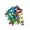
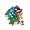
| Structure viewer | Molecule: MolmilJmol/JSmol |
|---|
- Downloads & links
-Download
| PDBx/mmCIF format | 1jn1.cif.gz | 99.5 KB | Display | PDBx/mmCIF format |
|---|---|---|---|---|
| PDB format | pdb1jn1.ent.gz | 77.7 KB | Display | PDB format |
| PDBx/mmJSON format | 1jn1.json.gz | Tree view | PDBx/mmJSON format | |
| Others |  Other downloads Other downloads |
-Validation report
| Arichive directory | https://data.pdbj.org/pub/pdb/validation_reports/jn/1jn1ftp://data.pdbj.org/pub/pdb/validation_reports/jn/1jn1 | HTTPS FTP |
|---|
-Related structure data
| Similar structure data | |
|---|---|
| Other databases |


-Links
 PDBj
PDBj
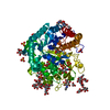
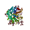
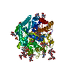
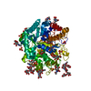
- Assembly
| Deposited unit | 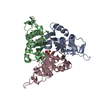
| ||||||||
|---|---|---|---|---|---|---|---|---|---|
| 1 |
| ||||||||
| 2 | 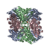
| ||||||||
| Unit cell |
| ||||||||
| Details | The protein is a trimer in solution. The asymmetric unit contains the homotrimer / A tetramer of trimers is formed in the crystal. The biological significance is unknown. To form a tetramer of trimers, use the following: X,Y,Z; 1-Y,1-X,1-Z; 1-X,1-Y,Z; Y,X,1-Z |
-Components
| #1: Protein | Mass: 17216.682 Da / Num. of mol.: 3 Source method: isolated from a genetically manipulated source Source: (gene. exp.) Haemophilus influenzae (bacteria) / Gene: ISPF / Plasmid: pET15b / Species (production host): Escherichia coli / Production host: #2: Chemical | ChemComp-CO /   Mass: 58.933 Da / Num. of mol.: 4 / Source method: obtained synthetically / Formula: Co Mass: 58.933 Da / Num. of mol.: 4 / Source method: obtained synthetically / Formula: Co#3: Chemical | ChemComp-SO4 / |   Mass: 96.063 Da / Num. of mol.: 1 / Source method: obtained synthetically / Formula: SO4 Mass: 96.063 Da / Num. of mol.: 1 / Source method: obtained synthetically / Formula: SO4 |
|---|
-Experimental details
-Experiment
| Experiment | Method: X-RAY DIFFRACTION / Number of used crystals: 1 |
|---|
- Sample preparation
| Crystal | Density Matthews: 2.86 Å3/Da / Density % sol: 57.02 % | |||||||||||||||||||||||||||||||||||
|---|---|---|---|---|---|---|---|---|---|---|---|---|---|---|---|---|---|---|---|---|---|---|---|---|---|---|---|---|---|---|---|---|---|---|---|---|
| Crystal grow | Temperature: 295 K / Method: vapor diffusion, hanging drop / pH: 6.5 Details: 40% sat. ammonium sulfate, 0.1 MES, 15 mM CoCl2, pH 6.5, VAPOR DIFFUSION, HANGING DROP, temperature 295K | |||||||||||||||||||||||||||||||||||
| Crystal grow | *PLUS | |||||||||||||||||||||||||||||||||||
| Components of the solutions | *PLUS
|
-Data collection
| Diffraction | Mean temperature: 100 K |
|---|---|
| Diffraction source | Source: SYNCHROTRON / Site: APS  / Beamline: 17-ID / Wavelength: 1 Å / Beamline: 17-ID / Wavelength: 1 Å |
| Detector | Type: MARRESEARCH / Detector: CCD / Date: Mar 30, 2001 |
| Radiation | Monochromator: Cryogenically-cooled Si(111) double-crystal system Protocol: SINGLE WAVELENGTH / Monochromatic (M) / Laue (L): M / Scattering type: x-ray |
| Radiation wavelength | Wavelength: 1 Å / Relative weight: 1 |
| Reflection | Resolution: 2.9→30 Å / Num. all: 13670 / Num. obs: 13670 / % possible obs: 99.9 % / Observed criterion σ(F): 0 / Observed criterion σ(I): 0 / Redundancy: 18.6 % / Rmerge(I) obs: 0.053 / Net I/σ(I): 9.4 |
| Reflection shell | Resolution: 2.9→3.03 Å / Rmerge(I) obs: 0.294 / % possible all: 100 |
| Reflection | *PLUS Highest resolution: 2.9 Å / Lowest resolution: 29 Å / % possible obs: 100 % / Num. measured all: 399292 / Rmerge(I) obs: 0.053 |
| Reflection shell | *PLUS % possible obs: 100 % / Rmerge(I) obs: 0.294 / Mean I/σ(I) obs: 9.4 |
- Processing
| Software |
| ||||||||||||||||||||
|---|---|---|---|---|---|---|---|---|---|---|---|---|---|---|---|---|---|---|---|---|---|
| Refinement | Method to determine structure: MOLECULAR REPLACEMENT Starting model: structure from MIR data in P6(3) space group Resolution: 2.9→20 Å / Cross valid method: THROUGHOUT / σ(F): 2 / Stereochemistry target values: engh & huber
| ||||||||||||||||||||
| Displacement parameters | Biso mean: 57 Å2 | ||||||||||||||||||||
| Refinement step | Cycle: LAST / Resolution: 2.9→20 Å
| ||||||||||||||||||||
| Refine LS restraints |
| ||||||||||||||||||||
| LS refinement shell | Resolution: 2.9→3 Å / Rfactor Rfree: 0.387 / Rfactor Rwork: 0.294 | ||||||||||||||||||||
| Refinement | *PLUS Highest resolution: 2.9 Å / σ(F): 2 / % reflection Rfree: 8 % / Rfactor Rfree: 0.29 / Rfactor Rwork: 0.198 | ||||||||||||||||||||
| Solvent computation | *PLUS | ||||||||||||||||||||
| Displacement parameters | *PLUS | ||||||||||||||||||||
| LS refinement shell | *PLUS Rfactor Rfree: 0.387 / Rfactor Rwork: 0.294 |
