 Movie
Movie Controller

[English] 日本語
 Yorodumi
- PDB-1j8b: Structure of YbaB from Haemophilus influenzae (HI0442), a protein... -
+ Open data

- Basic information
| Entry | Database: PDB / ID: 1j8b | ||||||
|---|---|---|---|---|---|---|---|
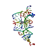
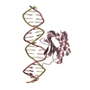
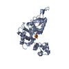
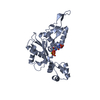
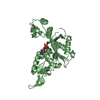
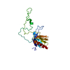
| Title | Structure of YbaB from Haemophilus influenzae (HI0442), a protein of unknown function | ||||||
 Components Components | YbaB | ||||||
 Keywords Keywords | STRUCTURAL GENOMICS / UNKNOWN FUNCTION / HI0442 / hypothetical protein / Structure 2 Function Project / S2F | ||||||
| Function / homology |  Function and homology information Function and homology information | ||||||
| Biological species |  Haemophilus influenzae Rd (bacteria) Haemophilus influenzae Rd (bacteria) | ||||||
| Method |  X-RAY DIFFRACTION / SYNCHROTRON / MAD / Resolution: 1.75 Å X-RAY DIFFRACTION / SYNCHROTRON / MAD / Resolution: 1.75 Å | ||||||
 Authors Authors | Lim, K. / Tempcyzk, A. / Toedt, J. / Parsons, J.F. / Howard, A. / Eisenstein, E. / Herzberg, O. / Structure 2 Function Project (S2F) | ||||||
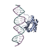
 Citation Citation | Journal: Proteins / Year: 2003 Title: Crystal structure of YbaB from Haemophilus influenzae (HI0442), a protein of unknown function coexpressed with the recombinational DNA repair protein RecR Authors: Lim, K. / Tempcyzk, A. / Parsons, J.F. / Bonander, N. / Toedt, J. / Kelman, Z. / Howard, A. / Eisenstein, E. / Herzberg, O. | ||||||
| History |
|
- Structure visualization
| Structure viewer | Molecule: MolmilJmol/JSmol |
|---|
- Downloads & links
-Download
| PDBx/mmCIF format | 1j8b.cif.gz | 31.7 KB | Display | PDBx/mmCIF format |
|---|---|---|---|---|
| PDB format | pdb1j8b.ent.gz | 24.6 KB | Display | PDB format |
| PDBx/mmJSON format | 1j8b.json.gz | Tree view | PDBx/mmJSON format | |
| Others |  Other downloads Other downloads |
-Validation report
| Summary document | 1j8b_validation.pdf.gz | 427.4 KB | Display | wwPDB validaton report |
|---|---|---|---|---|
| Full document | 1j8b_full_validation.pdf.gz | 429 KB | Display | |
| Data in XML | 1j8b_validation.xml.gz | 8.1 KB | Display | |
| Data in CIF | 1j8b_validation.cif.gz | 11.1 KB | Display | |
| Arichive directory | https://data.pdbj.org/pub/pdb/validation_reports/j8/1j8bftp://data.pdbj.org/pub/pdb/validation_reports/j8/1j8b | HTTPS FTP |
-Related structure data
| Similar structure data | |
|---|---|
| Other databases |

-Links
 PDBj
PDBj- Assembly
| Deposited unit | 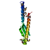
| |||||||||
|---|---|---|---|---|---|---|---|---|---|---|
| 1 | 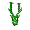
| |||||||||
| Unit cell |
| |||||||||
| Components on special symmetry positions |
| |||||||||
| Details | To obtain a dimer, apply -X,Y,1/2-Z symmetry operator and 1,0,1 translation vector |
-Components
| #1: Protein | Mass: 12734.129 Da / Num. of mol.: 1 Source method: isolated from a genetically manipulated source Source: (gene. exp.) Haemophilus influenzae Rd (bacteria) / Species: Haemophilus influenzae / Strain: KW20 / Gene: HI0442 / Plasmid: pET15b-HI0442 / Production host: |
|---|---|
| #2: Water | ChemComp-HOH /  Mass: 18.015 Da / Num. of mol.: 161 / Source method: isolated from a natural source / Formula: H2O Mass: 18.015 Da / Num. of mol.: 161 / Source method: isolated from a natural source / Formula: H2O |
-Experimental details
-Experiment
| Experiment | Method: X-RAY DIFFRACTION / Number of used crystals: 1 |
|---|
- Sample preparation
| Crystal | Density Matthews: 2.09 Å3/Da / Density % sol: 41.29 % | ||||||||||||||||||||||||||||||||||||
|---|---|---|---|---|---|---|---|---|---|---|---|---|---|---|---|---|---|---|---|---|---|---|---|---|---|---|---|---|---|---|---|---|---|---|---|---|---|
| Crystal grow | Temperature: 277 K / Method: vapor diffusion, hanging drop / pH: 3.8 Details: 1 M ammonium phosphate, pH 3.8, VAPOR DIFFUSION, HANGING DROP, temperature 277K | ||||||||||||||||||||||||||||||||||||
| Crystal grow | *PLUS pH: 7.5 | ||||||||||||||||||||||||||||||||||||
| Components of the solutions | *PLUS
|
-Data collection
| Diffraction | Mean temperature: 100 K | ||||||||||||
|---|---|---|---|---|---|---|---|---|---|---|---|---|---|
| Diffraction source | Source: SYNCHROTRON / Site: APS  / Beamline: 17-ID / Wavelength: 0.9789, 0.9790, 0.9500 / Beamline: 17-ID / Wavelength: 0.9789, 0.9790, 0.9500 | ||||||||||||
| Detector | Type: MARRESEARCH / Detector: CCD / Date: Jan 1, 2000 | ||||||||||||
| Radiation | Protocol: MAD / Monochromatic (M) / Laue (L): M / Scattering type: x-ray | ||||||||||||
| Radiation wavelength |
| ||||||||||||
| Reflection | Resolution: 1.75→99 Å / Num. all: 10936 / Num. obs: 10936 / % possible obs: 96.7 % / Observed criterion σ(F): 0 / Observed criterion σ(I): 0 / Redundancy: 6.5 % / Biso Wilson estimate: 21 Å2 / Rmerge(I) obs: 0.071 / Net I/σ(I): 18 | ||||||||||||
| Reflection shell | Resolution: 1.75→1.81 Å / Redundancy: 4.2 % / Rmerge(I) obs: 0.214 / Num. unique all: 810 / % possible all: 73.5 | ||||||||||||
| Reflection | *PLUS Lowest resolution: 99 Å / Num. obs: 19416 / Num. measured all: 73832 / Rmerge(I) obs: 0.046 | ||||||||||||
| Reflection shell | *PLUS % possible obs: 73.5 % / Rmerge(I) obs: 0.201 |
- Processing
| Software |
| |||||||||||||||||||||||||
|---|---|---|---|---|---|---|---|---|---|---|---|---|---|---|---|---|---|---|---|---|---|---|---|---|---|---|
| Refinement | Method to determine structure: MAD / Resolution: 1.75→99 Å / Cross valid method: THROUGHOUT / σ(F): 2 / Stereochemistry target values: Engh & Huber
| |||||||||||||||||||||||||
| Displacement parameters | Biso mean: 33 Å2 | |||||||||||||||||||||||||
| Refinement step | Cycle: LAST / Resolution: 1.75→99 Å
| |||||||||||||||||||||||||
| Refine LS restraints |
| |||||||||||||||||||||||||
| LS refinement shell | Resolution: 1.75→1.83 Å /
| |||||||||||||||||||||||||
| Refinement | *PLUS Lowest resolution: 20 Å / σ(F): 2 / Rfactor Rfree: 0.274 / Rfactor Rwork: 0.185 | |||||||||||||||||||||||||
| Solvent computation | *PLUS | |||||||||||||||||||||||||
| Displacement parameters | *PLUS | |||||||||||||||||||||||||
| LS refinement shell | *PLUS Rfactor Rfree: 0.331 / Rfactor Rwork: 0.353 |
