 Movie
Movie Controller

[English] 日本語
 Yorodumi
- PDB-1h0s: 3-dehydroquinate dehydratase from Mycobacterium tuberculosis in c... -
+ Open data

- Basic information
| Entry | Database: PDB / ID: 1h0s | ||||||
|---|---|---|---|---|---|---|---|
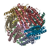
| Title | 3-dehydroquinate dehydratase from Mycobacterium tuberculosis in complex with 3-hydroxyimino-quinic acid | ||||||
 Components Components | 3-DEHYDROQUINATE DEHYDRATASE | ||||||
 Keywords Keywords | LYASE / SHIKIMATE PATHWAY / ALPHA/BETA PROTEIN | ||||||
| Function / homology |  Function and homology information Function and homology informationquinate catabolic process / Chorismate via Shikimate Pathway / 3-dehydroquinate dehydratase / 3-dehydroquinate dehydratase activity / chorismate biosynthetic process / aromatic amino acid family biosynthetic process / amino acid biosynthetic process / cytosolSimilarity search - Function | ||||||
| Biological species |   MYCOBACTERIUM TUBERCULOSIS (bacteria) MYCOBACTERIUM TUBERCULOSIS (bacteria) | ||||||
| Method | X-RAY DIFFRACTION / SYNCHROTRON / MOLECULAR REPLACEMENT / Resolution: 1.7 Å | ||||||
 Authors Authors | Roszak, A.W. / Frederickson, M. / Abell, C. / Coggins, J.R. / Lapthorn, A.J. | ||||||
 Citation Citation | Journal: To be Published Title: Structural Basis for Specificity of Oxime Based Inhibitors Towards Type II Dehydroquinase from Mycobacterium Tuberculosis Authors: Robinson, D.A. / Roszak, A.W. / Frederickson, M. / Abell, C. / Coggins, J.R. / Lapthorn, A.J. #1: Journal: Nat.Struct.Biol. / Year: 1999Title: The Two Types of 3-Dehydroquinase Have Distinct Structures But Catalyze the Same Overall Reaction. Authors: Gourley, D.G. / Shrive, A.K. / Polikarpov, I. / Krell, T. / Coggins, J.R. / Hawkins, A.R. / Isaacs, N.W. / Sawyer, L. | ||||||
| History |
|
- Structure visualization
| Structure viewer | Molecule: MolmilJmol/JSmol |
|---|
- Downloads & links
-Download
| PDBx/mmCIF format | 1h0s.cif.gz | 46.9 KB | Display | PDBx/mmCIF format |
|---|---|---|---|---|
| PDB format | pdb1h0s.ent.gz | 33.2 KB | Display | PDB format |
| PDBx/mmJSON format | 1h0s.json.gz | Tree view | PDBx/mmJSON format | |
| Others |  Other downloads Other downloads |
-Validation report
| Arichive directory | https://data.pdbj.org/pub/pdb/validation_reports/h0/1h0sftp://data.pdbj.org/pub/pdb/validation_reports/h0/1h0s | HTTPS FTP |
|---|
-Related structure data
| Related structure data |  2dhqS S: Starting model for refinement |
|---|---|
| Similar structure data |


-Links
 PDBj
PDBj
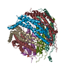
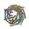
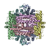
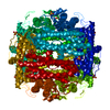
- Assembly
| Deposited unit | 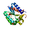
| |||||||||||||||||||||
|---|---|---|---|---|---|---|---|---|---|---|---|---|---|---|---|---|---|---|---|---|---|---|
| 1 | x 12
| |||||||||||||||||||||
| Unit cell |
| |||||||||||||||||||||
| Components on special symmetry positions |
|
-Components
| #1: Protein | / 3-DEHYDROQUINASE / TYPE II DHQASE Mass: 15676.737 Da / Num. of mol.: 1 Source method: isolated from a genetically manipulated source Source: (gene. exp.) MYCOBACTERIUM TUBERCULOSIS (bacteria) / Strain: H37RV / Plasmid: MPET / Production host: ESCHERICHIA COLI (E. coli) / Strain (production host): BL21(DE3)References: UniProt: P0A4Z6, UniProt: P9WPX7*PLUS, 3-dehydroquinate dehydratase | ||||
|---|---|---|---|---|---|
| #2: Chemical | ChemComp-FA6 / 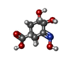  Mass: 205.165 Da / Num. of mol.: 1 / Source method: obtained synthetically / Formula: C7H11NO6 Mass: 205.165 Da / Num. of mol.: 1 / Source method: obtained synthetically / Formula: C7H11NO6 | ||||
| #3: Chemical | Sulfate  Mass: 96.063 Da / Num. of mol.: 3 / Source method: obtained synthetically / Formula: SO4 Mass: 96.063 Da / Num. of mol.: 3 / Source method: obtained synthetically / Formula: SO4#4: Chemical | Glycerol  Mass: 92.094 Da / Num. of mol.: 3 / Source method: obtained synthetically / Formula: C3H8O3 Mass: 92.094 Da / Num. of mol.: 3 / Source method: obtained synthetically / Formula: C3H8O3#5: Water | ChemComp-HOH / | Water Mass: 18.015 Da / Num. of mol.: 186 / Source method: isolated from a natural source / Formula: H2O Mass: 18.015 Da / Num. of mol.: 186 / Source method: isolated from a natural source / Formula: H2O |
-Experimental details
-Experiment
| Experiment | Method: X-RAY DIFFRACTION / Number of used crystals: 1 |
|---|
- Sample preparation
| Crystal | Density Matthews: 2.77 Å3/Da / Density % sol: 55.5 % |
|---|---|
| Crystal grow | pH: 6.5 Details: 20% ME2KPEG,0.5M AMMONIUM SULPHATE 0.1M MOPS PH 6.5 |
-Data collection
| Diffraction | Mean temperature: 100 K |
|---|---|
| Diffraction source | Source: SYNCHROTRON / Site: SRS  / Beamline: PX9.6 / Wavelength: 0.87 / Beamline: PX9.6 / Wavelength: 0.87 |
| Detector | Type: ADSC CCD / Detector: CCD / Date: Jul 7, 1999 |
| Radiation | Protocol: SINGLE WAVELENGTH / Monochromatic (M) / Laue (L): M / Scattering type: x-ray |
| Radiation wavelength | Wavelength: 0.87 Å / Relative weight: 1 |
| Reflection | Resolution: 1.7→70 Å / Num. obs: 117533 / % possible obs: 100 % / Redundancy: 6.3 % / Biso Wilson estimate: 21.62 Å2 / Rmerge(I) obs: 0.077 / Net I/σ(I): 12.3 |
| Reflection shell | Resolution: 1.69→1.73 Å / Redundancy: 3.6 % / Rmerge(I) obs: 0.808 / Mean I/σ(I) obs: 2.23 / % possible all: 100 |
- Processing
| Software |
| ||||||||||||||||||||
|---|---|---|---|---|---|---|---|---|---|---|---|---|---|---|---|---|---|---|---|---|---|
| Refinement | Method to determine structure: MOLECULAR REPLACEMENT Starting model: PDB ENTRY 2DHQ Resolution: 1.7→72.55 Å / SU B: 1.739 / SU ML: 0.056 / σ(F): 0 / ESU R: 0.087 / ESU R Free: 0.091 / Details: RESIDUES 20-25 ARE DISORDERED
| ||||||||||||||||||||
| Displacement parameters | Biso mean: 13.5 Å2 | ||||||||||||||||||||
| Refinement step | Cycle: LAST / Resolution: 1.7→72.55 Å
|
