 Movie
Movie Controller

[English] 日本語
 Yorodumi
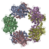
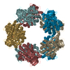
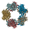
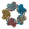
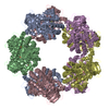
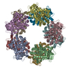
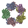
- PDB-1geh: CRYSTAL STRUCTURE OF ARCHAEAL RUBISCO (RIBULOSE 1,5-BISPHOSPHATE ... -
+ Open data

- Basic information
| Entry | Database: PDB / ID: 1geh | ||||||
|---|---|---|---|---|---|---|---|
| Title | CRYSTAL STRUCTURE OF ARCHAEAL RUBISCO (RIBULOSE 1,5-BISPHOSPHATE CARBOXYLASE/OXYGENASE) | ||||||
 Components Components | RIBULOSE-1,5-BISPHOSPHATE CARBOXYLASE/OXYGENASE | ||||||
 Keywords Keywords |  LYASE / Pentagonal toroid decamer / RUBISCO LYASE / Pentagonal toroid decamer / RUBISCO | ||||||
| Function / homology |  Function and homology information Function and homology informationAMP catabolic process / ribulose-bisphosphate carboxylase / carbon fixation / ribulose-bisphosphate carboxylase activity / oxidoreductase activity / magnesium ion bindingSimilarity search - Function | ||||||
| Biological species |   Thermococcus kodakarensis (archaea) Thermococcus kodakarensis (archaea) | ||||||
| Method | X-RAY DIFFRACTION / SYNCHROTRON / SIRAS / Resolution: 2.8 Å | ||||||
 Authors Authors | Kitano, K. / Maeda, N. / Fukui, T. / Atomi, H. / Imanaka, T. / Miki, K. | ||||||
 Citation Citation | Journal: Structure / Year: 2001 Title: Crystal Structure of a Novel-Type Archaeal Rubisco with Pentagonal Symmetry Authors: Kitano, K. / Maeda, N. / Fukui, T. / Atomi, H. / Imanaka, T. / Miki, K. #1: Journal: J.Mol.Biol. / Year: 1999Title: Ribulose Bisphosphate Carboxylase/oxygenase from Hyperthermophilic Archaeon Pyrococcus kodakaraensis KOD1 is Composed Solely of Large Subunits and Forms a Pentagonal Structure Authors: Maeda, N. / Kitano, K. / Fukui, T. / Ezaki, S. / Atomi, H. / Miki, K. / Imanaka, T. | ||||||
| History |
|
- Structure visualization
| Structure viewer | Molecule: MolmilJmol/JSmol |
|---|
- Downloads & links
-Download
| PDBx/mmCIF format | 1geh.cif.gz | 376.3 KB | Display | PDBx/mmCIF format |
|---|---|---|---|---|
| PDB format | pdb1geh.ent.gz | 316.6 KB | Display | PDB format |
| PDBx/mmJSON format | 1geh.json.gz | Tree view | PDBx/mmJSON format | |
| Others |  Other downloads Other downloads |
-Validation report
| Arichive directory | https://data.pdbj.org/pub/pdb/validation_reports/ge/1gehftp://data.pdbj.org/pub/pdb/validation_reports/ge/1geh | HTTPS FTP |
|---|
-Related structure data
| Similar structure data |
|---|

-Links
 PDBj
PDBj
- Assembly
| Deposited unit | 
| ||||||||
|---|---|---|---|---|---|---|---|---|---|
| 1 | 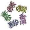
| ||||||||
| Unit cell |
| ||||||||
| Details | The biological assembly is a decamer generated from the pentamer in the asymmetric unit by the operations: y, x, -z. |
-Components
| #1: Protein | Mass: 49776.480 Da / Num. of mol.: 5 Source method: isolated from a genetically manipulated source Source: (gene. exp.) Thermococcus kodakarensis (archaea) / Strain: KOD1 / Production host:  Escherichia coli (E. coli) Escherichia coli (E. coli)References: UniProt: O93627, ribulose-bisphosphate carboxylase#2: Chemical | ChemComp-SO4 / Sulfate  Mass: 96.063 Da / Num. of mol.: 10 / Source method: obtained synthetically / Formula: SO4 Mass: 96.063 Da / Num. of mol.: 10 / Source method: obtained synthetically / Formula: SO4 |
|---|
-Experimental details
-Experiment
| Experiment | Method: X-RAY DIFFRACTION / Number of used crystals: 5 |
|---|
- Sample preparation
| Crystal | Density Matthews: 2.95 Å3/Da / Density % sol: 58.36 % | |||||||||||||||||||||||||||||||||||
|---|---|---|---|---|---|---|---|---|---|---|---|---|---|---|---|---|---|---|---|---|---|---|---|---|---|---|---|---|---|---|---|---|---|---|---|---|
| Crystal grow | Temperature: 293 K / Method: vapor diffusion / pH: 9 Details: Ammoinum sulfate, pH 9.0, VAPOR DIFFUSION, temperature 293K | |||||||||||||||||||||||||||||||||||
| Crystal grow | *PLUS Temperature: 20 ℃ / Method: vapor diffusion, hanging drop / Details: Maeda, N., (1999) J.Mol.Biol., 293, 57. | |||||||||||||||||||||||||||||||||||
| Components of the solutions | *PLUS
|
-Data collection
| Diffraction |
| |||||||||||||||
|---|---|---|---|---|---|---|---|---|---|---|---|---|---|---|---|---|
| Diffraction source |
| |||||||||||||||
| Detector |
| |||||||||||||||
| Radiation | Protocol: SINGLE WAVELENGTH / Monochromatic (M) / Laue (L): M / Scattering type: x-ray | |||||||||||||||
| Radiation wavelength | Wavelength: 1 Å / Relative weight: 1 | |||||||||||||||
| Reflection | Resolution: 2.8→100 Å / Num. obs: 64453 / % possible obs: 89.5 % / Observed criterion σ(F): 1 / Observed criterion σ(I): 1 / Redundancy: 12.1 % / Biso Wilson estimate: 17.3 Å2 / Rmerge(I) obs: 0.091 / Net I/σ(I): 11.8 | |||||||||||||||
| Reflection shell | Resolution: 2.8→2.9 Å / Rmerge(I) obs: 0.335 / % possible all: 65.5 | |||||||||||||||
| Reflection | *PLUS Lowest resolution: 100 Å / Num. measured all: 784332 | |||||||||||||||
| Reflection shell | *PLUS % possible obs: 65.5 % / Mean I/σ(I) obs: 3.4 |

- Processing
| Software |
| ||||||||||||||||||||||||||||||||||||||||
|---|---|---|---|---|---|---|---|---|---|---|---|---|---|---|---|---|---|---|---|---|---|---|---|---|---|---|---|---|---|---|---|---|---|---|---|---|---|---|---|---|---|
| Refinement | Method to determine structure: SIRAS / Resolution: 2.8→100 Å / Rfactor Rfree error: 0.003 / Data cutoff high absF: 11437229 / Data cutoff low absF: 0 / σ(F): 1 / σ(I): 1 / Stereochemistry target values: Engh & Huber
| ||||||||||||||||||||||||||||||||||||||||
| Solvent computation | Solvent model: FLAT MODEL | ||||||||||||||||||||||||||||||||||||||||
| Refinement step | Cycle: LAST / Resolution: 2.8→100 Å
| ||||||||||||||||||||||||||||||||||||||||
| Refine LS restraints |
| ||||||||||||||||||||||||||||||||||||||||
| LS refinement shell | Highest resolution: 2.8 Å / Total num. of bins used: 6 /
| ||||||||||||||||||||||||||||||||||||||||
| Xplor file |
| ||||||||||||||||||||||||||||||||||||||||
| Software | *PLUS Name: CNS / Version: 1 / Classification: refinement | ||||||||||||||||||||||||||||||||||||||||
| Refinement | *PLUS Highest resolution: 2.8 Å / Lowest resolution: 100 Å / σ(F): 1 / % reflection Rfree: 10.1 % / Rfactor obs: 0.224 | ||||||||||||||||||||||||||||||||||||||||
| Solvent computation | *PLUS | ||||||||||||||||||||||||||||||||||||||||
| Displacement parameters | *PLUS | ||||||||||||||||||||||||||||||||||||||||
| Refine LS restraints | *PLUS
| ||||||||||||||||||||||||||||||||||||||||
| LS refinement shell | *PLUS % reflection Rfree: 10.1 % |
