 Movie
Movie Controller

[English] 日本語
 Yorodumi
- PDB-1ga6: CRYSTAL STRUCTURE ANALYSIS OF PSCP (PSEUDOMONAS SERINE-CARBOXYL P... -
+ Open data

- Basic information
| Entry | Database: PDB / ID: 1ga6 | ||||||
|---|---|---|---|---|---|---|---|
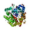
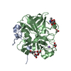
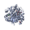
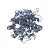
| Title | CRYSTAL STRUCTURE ANALYSIS OF PSCP (PSEUDOMONAS SERINE-CARBOXYL PROTEINASE) COMPLEXED WITH A FRAGMENT OF TYROSTATIN (THIS ENZYME RENAMED "SEDOLISIN" IN 2003) | ||||||
 Components Components |
| ||||||
 Keywords Keywords | HYDROLASE/HYDROLASE INHIBITOR / SERINE-CARBOXYL PROTEINASE / HYDROLASE-HYDROLASE INHIBITOR complex | ||||||
| Function / homology |  Function and homology information Function and homology informationsedolisin / tripeptidyl-peptidase activity / periplasmic space / serine-type endopeptidase activity / proteolysis / metal ion binding Similarity search - Function | ||||||
| Biological species |  Pseudomonas sp. (bacteria) Pseudomonas sp. (bacteria) | ||||||
| Method |  X-RAY DIFFRACTION / SYNCHROTRON / ISOMORPHOUS WITH 1GA1 / Resolution: 1 Å X-RAY DIFFRACTION / SYNCHROTRON / ISOMORPHOUS WITH 1GA1 / Resolution: 1 Å | ||||||
 Authors Authors | Wlodawer, A. / Li, M. / Dauter, Z. / Gustchina, A. / Uchida, K. | ||||||
 Citation Citation | Journal: Nat.Struct.Biol. / Year: 2001 Title: Carboxyl proteinase from Pseudomonas defines a novel family of subtilisin-like enzymes. Authors: Wlodawer, A. / Li, M. / Dauter, Z. / Gustchina, A. / Uchida, K. / Oyama, H. / Dunn, B.M. / Oda, K. | ||||||
| History |
|
- Structure visualization
| Structure viewer | Molecule: MolmilJmol/JSmol |
|---|
- Downloads & links
-Download
| PDBx/mmCIF format | 1ga6.cif.gz | 174.7 KB | Display | PDBx/mmCIF format |
|---|---|---|---|---|
| PDB format | pdb1ga6.ent.gz | 137 KB | Display | PDB format |
| PDBx/mmJSON format | 1ga6.json.gz | Tree view | PDBx/mmJSON format | |
| Others |  Other downloads Other downloads |
-Validation report
| Summary document | 1ga6_validation.pdf.gz | 446.2 KB | Display | wwPDB validaton report |
|---|---|---|---|---|
| Full document | 1ga6_full_validation.pdf.gz | 450.8 KB | Display | |
| Data in XML | 1ga6_validation.xml.gz | 20.8 KB | Display | |
| Data in CIF | 1ga6_validation.cif.gz | 32.8 KB | Display | |
| Arichive directory | https://data.pdbj.org/pub/pdb/validation_reports/ga/1ga6ftp://data.pdbj.org/pub/pdb/validation_reports/ga/1ga6 | HTTPS FTP |
-Related structure data
| Related structure data |  1ga4C  1ga1S S: Starting model for refinement C: citing same article ( |
|---|---|
| Similar structure data |

-Links
 PDBj
PDBj

- Assembly
| Deposited unit | 
| |||||||||
|---|---|---|---|---|---|---|---|---|---|---|
| 1 |
| |||||||||
| Unit cell |
| |||||||||
| Components on special symmetry positions |
|
-Components
-Protein / Protein/peptide , 2 types, 2 molecules AI
| #1: Protein | Mass: 38446.191 Da / Num. of mol.: 1 Source method: isolated from a genetically manipulated source Source: (gene. exp.) Pseudomonas sp. (bacteria) / Plasmid: PUKCP2212 / Production host: |
|---|---|
| #2: Protein/peptide | Mass: 351.399 Da / Num. of mol.: 1 / Source method: obtained synthetically / Details: The inhibitor was chemically synthesized. |
-Non-polymers , 4 types, 475 molecules 


| #3: Chemical | ChemComp-CA /  Mass: 40.078 Da / Num. of mol.: 1 / Source method: obtained synthetically / Formula: Ca Mass: 40.078 Da / Num. of mol.: 1 / Source method: obtained synthetically / Formula: Ca | ||||
|---|---|---|---|---|---|
| #4: Chemical | ChemComp-ACT /  Mass: 59.044 Da / Num. of mol.: 4 / Source method: obtained synthetically / Formula: C2H3O2 Mass: 59.044 Da / Num. of mol.: 4 / Source method: obtained synthetically / Formula: C2H3O2#5: Chemical | ChemComp-GOL /  Mass: 92.094 Da / Num. of mol.: 4 / Source method: obtained synthetically / Formula: C3H8O3 Mass: 92.094 Da / Num. of mol.: 4 / Source method: obtained synthetically / Formula: C3H8O3#6: Water | ChemComp-HOH / | Mass: 18.015 Da / Num. of mol.: 466 / Source method: isolated from a natural source / Formula: H2O |
-Details
| Compound details | TYROSTATIN IS A NATURAL INHIBITOR, COMPOSED OF ISOVALERYL-TYROSYL-LEUSYL-TYROSINAL. IN 1GA1 AND ...TYROSTATIN |
|---|
-Experimental details
-Experiment
| Experiment | Method: X-RAY DIFFRACTION / Number of used crystals: 1 |
|---|
- Sample preparation
| Crystal | Density Matthews: 2.94 Å3/Da / Density % sol: 58.17 % | |||||||||||||||||||||||||||||||||||
|---|---|---|---|---|---|---|---|---|---|---|---|---|---|---|---|---|---|---|---|---|---|---|---|---|---|---|---|---|---|---|---|---|---|---|---|---|
| Crystal grow | Temperature: 293 K / Method: vapor diffusion, hanging drop / pH: 5.2 Details: 5% guanidine, 7% glycerol, 5% methanol, 1.0 M ammonium sulfate, 0.1 M sodium citrate buffer pH 5.2, VAPOR DIFFUSION, HANGING DROP, temperature 293K | |||||||||||||||||||||||||||||||||||
| Crystal grow | *PLUS pH: 4.8 | |||||||||||||||||||||||||||||||||||
| Components of the solutions | *PLUS
|
-Data collection
| Diffraction | Mean temperature: 100 K |
|---|---|
| Diffraction source | Source: SYNCHROTRON / Site: NSLS  / Beamline: X9B / Wavelength: 0.98 Å / Beamline: X9B / Wavelength: 0.98 Å |
| Detector | Type: ADSC QUANTUM 4 / Detector: CCD / Date: Aug 3, 2000 / Details: focussing mirror |
| Radiation | Monochromator: Si(111) double crystal, sagitally focussing / Protocol: SINGLE WAVELENGTH / Monochromatic (M) / Laue (L): M / Scattering type: x-ray |
| Radiation wavelength | Wavelength: 0.98 Å / Relative weight: 1 |
| Reflection | Resolution: 1→10 Å / Num. all: 241182 / Num. obs: 202008 / % possible obs: 84 % / Observed criterion σ(F): -3 / Observed criterion σ(I): -3 / Redundancy: 10.6 % / Biso Wilson estimate: 6.58 Å2 / Rmerge(I) obs: 0.054 / Net I/σ(I): 38.7 |
| Reflection shell | Resolution: 1→1.04 Å / Redundancy: 10.6 % / Rmerge(I) obs: 0.477 / Mean I/σ(I) obs: 4.7 / Num. unique all: 24057 / % possible all: 10 |
| Reflection | *PLUS Num. measured all: 2554920 |
| Reflection shell | *PLUS % possible obs: 100 % |
- Processing
| Software |
| |||||||||||||||||||||||||
|---|---|---|---|---|---|---|---|---|---|---|---|---|---|---|---|---|---|---|---|---|---|---|---|---|---|---|
| Refinement | Method to determine structure: ISOMORPHOUS WITH 1GA1 Starting model: PDB ENTRY 1GA1 Resolution: 1→10 Å / Cross valid method: R-FREE / σ(F): -3 / σ(I): -3 / Stereochemistry target values: Engh & Huber Details: Anisotropic refinement of individual atoms. Parameter errors estimated from least-squares blocked full-matrix inversion.
| |||||||||||||||||||||||||
| Refinement step | Cycle: LAST / Resolution: 1→10 Å
| |||||||||||||||||||||||||
| Refine LS restraints |
| |||||||||||||||||||||||||
| LS refinement shell | Resolution: 1→10 Å
|
