 Movie
Movie Controller


+ Open data

- Basic information
| Entry | Database: PDB / ID: 1g2c | ||||||
|---|---|---|---|---|---|---|---|
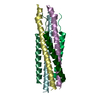
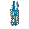
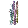
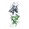
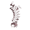
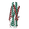
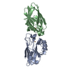
| Title | HUMAN RESPIRATORY SYNCYTIAL VIRUS FUSION PROTEIN CORE | ||||||
 Components Components | (FUSION PROTEIN (F)) x 2 | ||||||
 Keywords Keywords | VIRAL PROTEIN / membrane fusion / pneumovirus / HRSV | ||||||
| Function / homology |  Function and homology information Function and homology informationsymbiont-mediated induction of syncytium formation / host cell Golgi membrane / entry receptor-mediated virion attachment to host cell / fusion of virus membrane with host plasma membrane / viral envelope / symbiont entry into host cell / host cell plasma membrane / virion membrane / membrane Similarity search - Function | ||||||
| Biological species |  Human respiratory syncytial virus Human respiratory syncytial virus | ||||||
| Method |  X-RAY DIFFRACTION / SYNCHROTRON / Resolution: 2.3 Å X-RAY DIFFRACTION / SYNCHROTRON / Resolution: 2.3 Å | ||||||
 Authors Authors | Zhao, X. / Singh, M. / Malashkevich, V.N. / Kim, P.S. | ||||||
 Citation Citation | Journal: Proc.Natl.Acad.Sci.USA / Year: 2000 Title: Structural characterization of the human respiratory syncytial virus fusion protein core. Authors: Zhao, X. / Singh, M. / Malashkevich, V.N. / Kim, P.S. | ||||||
| History |
|
- Structure visualization
| Structure viewer | Molecule: MolmilJmol/JSmol |
|---|
- Downloads & links
-Download
| PDBx/mmCIF format | 1g2c.cif.gz | 221.8 KB | Display | PDBx/mmCIF format |
|---|---|---|---|---|
| PDB format | pdb1g2c.ent.gz | 182.4 KB | Display | PDB format |
| PDBx/mmJSON format | 1g2c.json.gz | Tree view | PDBx/mmJSON format | |
| Others |  Other downloads Other downloads |
-Validation report
| Arichive directory | https://data.pdbj.org/pub/pdb/validation_reports/g2/1g2cftp://data.pdbj.org/pub/pdb/validation_reports/g2/1g2c | HTTPS FTP |
|---|
-Related structure data
| Similar structure data |
|---|


-Links
 PDBj
PDBj

- Assembly
| Deposited unit | 
| ||||||||||
|---|---|---|---|---|---|---|---|---|---|---|---|
| 1 | 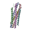
| ||||||||||
| 2 | 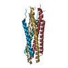
| ||||||||||
| 3 | 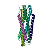
| ||||||||||
| 4 | 
| ||||||||||
| Unit cell |
| ||||||||||
| Details | The biological assembly is a trimer of heterodimer constructed from chain A, B, C,D,E,F |
-Components
| #1: Protein | Mass: 5653.654 Da / Num. of mol.: 12 / Fragment: RESIDUES 153-209, HRSV F1 HEPTAD REPEAT Source method: isolated from a genetically manipulated source Details: FIRST HEPTAD REPEAT / Source: (gene. exp.) Human respiratory syncytial virus / Genus: Pneumovirus / Strain: RSS-2 / Gene: HRSV F / Plasmid: PQE9 / Production host:  #2: Protein/peptide | Mass: 4873.347 Da / Num. of mol.: 12 / Fragment: RESIDUES 476-520, HRSV F1 HEPTAD REPEAT Source method: isolated from a genetically manipulated source Details: SECOND HEPTAD REPEAT / Source: (gene. exp.) Human respiratory syncytial virus / Genus: Pneumovirus / Strain: RSS-2 / Gene: HRSV F / Plasmid: PQE9 / Production host: #3: Water | ChemComp-HOH / |  Mass: 18.015 Da / Num. of mol.: 888 / Source method: isolated from a natural source / Formula: H2O Mass: 18.015 Da / Num. of mol.: 888 / Source method: isolated from a natural source / Formula: H2O |
|---|
-Experimental details
-Experiment
| Experiment | Method: X-RAY DIFFRACTION / Number of used crystals: 1 |
|---|
- Sample preparation
| Crystal | Density Matthews: 2.28 Å3/Da / Density % sol: 46.9 % | ||||||||||||||||||||||||||||||
|---|---|---|---|---|---|---|---|---|---|---|---|---|---|---|---|---|---|---|---|---|---|---|---|---|---|---|---|---|---|---|---|
| Crystal grow | Temperature: 293 K / Method: vapor diffusion, hanging drop / pH: 8.5 Details: PEG 4000, Tris-HCl, Li2SO4, pH 8.5, VAPOR DIFFUSION, HANGING DROP, temperature 293K | ||||||||||||||||||||||||||||||
| Crystal grow | *PLUS | ||||||||||||||||||||||||||||||
| Components of the solutions | *PLUS
|
-Data collection
| Diffraction | Mean temperature: 100 K |
|---|---|
| Diffraction source | Source: SYNCHROTRON / Site: NSLS  / Beamline: X4A / Wavelength: 0.9816 Å / Beamline: X4A / Wavelength: 0.9816 Å |
| Detector | Type: ADSC QUANTUM 4 / Detector: CCD / Date: Jul 20, 1999 |
| Radiation | Protocol: SINGLE WAVELENGTH / Monochromatic (M) / Laue (L): M / Scattering type: x-ray |
| Radiation wavelength | Wavelength: 0.9816 Å / Relative weight: 1 |
| Reflection | Resolution: 2.2→35 Å / Num. all: 62470 / Num. obs: 56973 / % possible obs: 91.2 % / Observed criterion σ(I): -3 / Redundancy: 1.3 % / Biso Wilson estimate: 40.37 Å2 / Rmerge(I) obs: 0.036 / Net I/σ(I): 8.9 |
| Reflection shell | Resolution: 2.2→2.28 Å / Redundancy: 2 % / Rmerge(I) obs: 0.24 / Num. unique all: 6224 / % possible all: 64.7 |
| Reflection | *PLUS Num. measured all: 119905 |
| Reflection shell | *PLUS % possible obs: 64.7 % |
- Processing
| Software |
| ||||||||||||||||||||||||
|---|---|---|---|---|---|---|---|---|---|---|---|---|---|---|---|---|---|---|---|---|---|---|---|---|---|
| Refinement | Starting model: modified polyser SV5 Resolution: 2.3→10 Å / Rfactor Rfree error: 0.004 / Data cutoff high absF: 1137175.53 / Data cutoff low absF: 0 / Isotropic thermal model: RESTRAINED / Cross valid method: THROUGHOUT / σ(F): 0 / Stereochemistry target values: MLF
| ||||||||||||||||||||||||
| Solvent computation | Solvent model: FLAT MODEL / Bsol: 56.95 Å2 / ksol: 0.283 e/Å3 | ||||||||||||||||||||||||
| Displacement parameters | Biso mean: 54.6 Å2
| ||||||||||||||||||||||||
| Refine analyze |
| ||||||||||||||||||||||||
| Refinement step | Cycle: LAST / Resolution: 2.3→10 Å
| ||||||||||||||||||||||||
| Refine LS restraints |
| ||||||||||||||||||||||||
| LS refinement shell | Resolution: 2.3→2.44 Å / Rfactor Rfree error: 0.012 / Total num. of bins used: 6
| ||||||||||||||||||||||||
| Xplor file |
| ||||||||||||||||||||||||
| Software | *PLUS Name: CNS / Version: 0.5 / Classification: refinement | ||||||||||||||||||||||||
| Refinement | *PLUS σ(F): 0 / % reflection Rfree: 10.2 % | ||||||||||||||||||||||||
| Solvent computation | *PLUS | ||||||||||||||||||||||||
| Displacement parameters | *PLUS Biso mean: 54.6 Å2 | ||||||||||||||||||||||||
| Refine LS restraints | *PLUS
| ||||||||||||||||||||||||
| LS refinement shell | *PLUS Rfactor Rfree: 0.352 / % reflection Rfree: 10.1 % / Rfactor Rwork: 0.292 |
