 Movie
Movie Controller

[English] 日本語
 Yorodumi
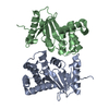
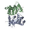
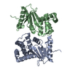
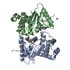
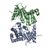
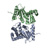
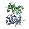
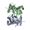
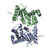
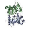
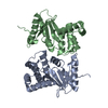
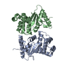
- PDB-1fvk: THE 1.7 ANGSTROM STRUCTURE OF WILD TYPE DISULFIDE BOND FORMATION ... -
+ Open data

- Basic information
| Entry | Database: PDB / ID: 1fvk | ||||||
|---|---|---|---|---|---|---|---|
| Title | THE 1.7 ANGSTROM STRUCTURE OF WILD TYPE DISULFIDE BOND FORMATION PROTEIN (DSBA) | ||||||
 Components Components | DISULFIDE BOND FORMATION PROTEIN | ||||||
 Keywords Keywords | DISULFIDE OXIDOREDUCTASE / PROTEIN DISULFIDE ISOMERASE / PROTEIN FOLDING / REDOX PROTEIN / REDOX-ACTIVE CENTER | ||||||
| Function / homology |  Function and homology information Function and homology informationcellular response to antibiotic / protein disulfide isomerase activity / protein-disulfide reductase activity / outer membrane-bounded periplasmic space / periplasmic space / oxidoreductase activity Similarity search - Function | ||||||
| Biological species |  | ||||||
| Method |  X-RAY DIFFRACTION / MIR / Resolution: 1.7 Å X-RAY DIFFRACTION / MIR / Resolution: 1.7 Å | ||||||
 Authors Authors | Martin, J.L. / Guddat, L.W. | ||||||
 Citation Citation | Journal: Protein Sci. / Year: 1997 Title: Structural analysis of three His32 mutants of DsbA: support for an electrostatic role of His32 in DsbA stability. Authors: Guddat, L.W. / Bardwell, J.C. / Glockshuber, R. / Huber-Wunderlich, M. / Zander, T. / Martin, J.L. #1: Journal: Nature / Year: 1993Title: Crystal Structure of the Dsba Protein Required for Disulphide Bond Formation in Vivo Authors: Martin, J.L. / Bardwell, J.C. / Kuriyan, J. #2: Journal: J.Mol.Biol. / Year: 1993Title: Crystallization of Dsba, an Escherichia Coli Protein Required for Disulphide Bond Formation in Vivo Authors: Martin, J.L. / Waksman, G. / Bardwell, J.C. / Beckwith, J. / Kuriyan, J. | ||||||
| History |
| ||||||
| Remark 650 | HELIX HELIX A1' IS SEPARATED FROM A1 BY A THREE RESIDUE LOOP. HELIX B1' IS SEPARATED FROM B1 BY A ...HELIX HELIX A1' IS SEPARATED FROM A1 BY A THREE RESIDUE LOOP. HELIX B1' IS SEPARATED FROM B1 BY A THREE RESIDUE LOOP. HELIX A3 IS KINKED BY PRO A 91 AND HELIX B3 BY PRO B 91. |
- Structure visualization
| Structure viewer | Molecule: MolmilJmol/JSmol |
|---|
- Downloads & links
-Download
| PDBx/mmCIF format | 1fvk.cif.gz | 84.8 KB | Display | PDBx/mmCIF format |
|---|---|---|---|---|
| PDB format | pdb1fvk.ent.gz | 67.7 KB | Display | PDB format |
| PDBx/mmJSON format | 1fvk.json.gz | Tree view | PDBx/mmJSON format | |
| Others |  Other downloads Other downloads |
-Validation report
| Summary document | 1fvk_validation.pdf.gz | 433.7 KB | Display | wwPDB validaton report |
|---|---|---|---|---|
| Full document | 1fvk_full_validation.pdf.gz | 435.5 KB | Display | |
| Data in XML | 1fvk_validation.xml.gz | 17.8 KB | Display | |
| Data in CIF | 1fvk_validation.cif.gz | 25.5 KB | Display | |
| Arichive directory | https://data.pdbj.org/pub/pdb/validation_reports/fv/1fvkftp://data.pdbj.org/pub/pdb/validation_reports/fv/1fvk | HTTPS FTP |
-Related structure data
| Related structure data | 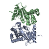 1ac1C 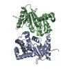 1acvC 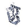 1fvjC  1dsdS S: Starting model for refinement C: citing same article ( |
|---|---|
| Similar structure data |


-Links
 PDBj
PDBj

- Assembly
| Deposited unit | 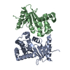
| ||||||||
|---|---|---|---|---|---|---|---|---|---|
| 1 |
| ||||||||
| Unit cell |
| ||||||||
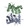
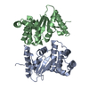
| Details | THERE ARE TWO MOLECULES IN THE ASYMMETRIC UNIT. EACH CONTAINS 189 RESIDUES, BUT THE LAST RESIDUE (189) IS NOT. OBSERVED. THE ACTIVE SITE DISULFIDE RESIDUES ARE CYS 30 AND CYS 33. PRO 151 FORMS PART OF THE ACTIVE SITE. |
-Components
| #1: Protein | Mass: 21155.025 Da / Num. of mol.: 2 / Source method: isolated from a natural source / Source: (natural) #2: Water | ChemComp-HOH / |  Mass: 18.015 Da / Num. of mol.: 248 / Source method: isolated from a natural source / Formula: H2O Mass: 18.015 Da / Num. of mol.: 248 / Source method: isolated from a natural source / Formula: H2O |
|---|
-Experimental details
-Experiment
| Experiment | Method: X-RAY DIFFRACTION / Number of used crystals: 1 |
|---|
- Sample preparation
| Crystal | Density Matthews: 2.79 Å3/Da / Density % sol: 55.86 % |
|---|---|
| Crystal grow | pH: 7.5 / Details: pH 7.5 |
-Data collection
| Diffraction | Mean temperature: 289 K |
|---|---|
| Diffraction source | Source: ROTATING ANODE / Type: RIGAKU RUH2R / Wavelength: 1.5418 |
| Detector | Type: RIGAKU / Detector: IMAGE PLATE / Date: Jul 10, 1995 / Details: YALE MIRRORS |
| Radiation | Monochromator: GRAPHITE(002) / Monochromatic (M) / Laue (L): M / Scattering type: x-ray |
| Radiation wavelength | Wavelength: 1.5418 Å / Relative weight: 1 |
| Reflection | Resolution: 1.7→50 Å / Num. obs: 46502 / % possible obs: 90.5 % / Observed criterion σ(I): 0.3 / Redundancy: 2.8 % / Biso Wilson estimate: 21 Å2 / Rmerge(I) obs: 0.0635 / Net I/σ(I): 14.1 |
| Reflection shell | Resolution: 1.7→1.78 Å / Redundancy: 1.7 % / Rmerge(I) obs: 0.328 / Mean I/σ(I) obs: 2.36 / % possible all: 84.1 |
- Processing
| Software |
| ||||||||||||||||||||||||||||||||||||||||||||||||||||||||||||||||||||||||||||||||
|---|---|---|---|---|---|---|---|---|---|---|---|---|---|---|---|---|---|---|---|---|---|---|---|---|---|---|---|---|---|---|---|---|---|---|---|---|---|---|---|---|---|---|---|---|---|---|---|---|---|---|---|---|---|---|---|---|---|---|---|---|---|---|---|---|---|---|---|---|---|---|---|---|---|---|---|---|---|---|---|---|---|
| Refinement | Method to determine structure: MIR Starting model: PDB ENTRY 1DSD Resolution: 1.7→50 Å / Cross valid method: YES / σ(F): 1
| ||||||||||||||||||||||||||||||||||||||||||||||||||||||||||||||||||||||||||||||||
| Displacement parameters | Biso mean: 33.1 Å2 | ||||||||||||||||||||||||||||||||||||||||||||||||||||||||||||||||||||||||||||||||
| Refine analyze |
| ||||||||||||||||||||||||||||||||||||||||||||||||||||||||||||||||||||||||||||||||
| Refinement step | Cycle: LAST / Resolution: 1.7→50 Å
| ||||||||||||||||||||||||||||||||||||||||||||||||||||||||||||||||||||||||||||||||
| Refine LS restraints |
| ||||||||||||||||||||||||||||||||||||||||||||||||||||||||||||||||||||||||||||||||
| LS refinement shell | Resolution: 1.7→1.78 Å
|
