 Movie
Movie Controller


+ Open data

- Basic information
| Entry | Database: PDB / ID: 1fm2 | ||||||
|---|---|---|---|---|---|---|---|
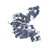
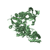
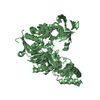
| Title | THE 2 ANGSTROM CRYSTAL STRUCTURE OF CEPHALOSPORIN ACYLASE | ||||||
 Components Components | (GLUTARYL 7-AMINOCEPHALOSPORANIC ACID ACYLASE) x 2 | ||||||
 Keywords Keywords | HYDROLASE / cephalosporin acylase / antibiotics / penicillin acylase / N-terminal hydrolase | ||||||
| Function / homology |  Function and homology information Function and homology informationglutaryl-7-aminocephalosporanic-acid acylase / glutaryl-7-aminocephalosporanic-acid acylase activity / antibiotic biosynthetic process / periplasmic space / response to antibiotic Similarity search - Function | ||||||
| Biological species |  Brevundimonas diminuta (bacteria) Brevundimonas diminuta (bacteria) | ||||||
| Method |  X-RAY DIFFRACTION / SYNCHROTRON / Resolution: 2 Å X-RAY DIFFRACTION / SYNCHROTRON / Resolution: 2 Å | ||||||
 Authors Authors | Kim, Y. / Yoon, K.H. / Khang, Y. / Turley, S. / Hol, W.G.J. | ||||||
 Citation Citation | Journal: Structure Fold.Des. / Year: 2000 Title: The 2.0 A crystal structure of cephalosporin acylase. Authors: Kim, Y. / Yoon, K. / Khang, Y. / Turley, S. / Hol, W.G. | ||||||
| History |
|
- Structure visualization
| Structure viewer | Molecule: MolmilJmol/JSmol |
|---|
- Downloads & links
-Download
| PDBx/mmCIF format | 1fm2.cif.gz | 154.2 KB | Display | PDBx/mmCIF format |
|---|---|---|---|---|
| PDB format | pdb1fm2.ent.gz | 118.7 KB | Display | PDB format |
| PDBx/mmJSON format | 1fm2.json.gz | Tree view | PDBx/mmJSON format | |
| Others |  Other downloads Other downloads |
-Validation report
| Arichive directory | https://data.pdbj.org/pub/pdb/validation_reports/fm/1fm2ftp://data.pdbj.org/pub/pdb/validation_reports/fm/1fm2 | HTTPS FTP |
|---|
-Related structure data
| Similar structure data |
|---|


-Links
 PDBj
PDBj


- Assembly
| Deposited unit | 
| ||||||||
|---|---|---|---|---|---|---|---|---|---|
| 1 |
| ||||||||
| Unit cell |
|
-Components
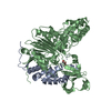
| #1: Protein | Mass: 18567.113 Da / Num. of mol.: 1 / Fragment: ALPHA SUBUNIT Source method: isolated from a genetically manipulated source Source: (gene. exp.) Brevundimonas diminuta (bacteria) / Plasmid: PET SYSTEM / Production host: |
|---|---|
| #2: Protein | Mass: 58686.363 Da / Num. of mol.: 1 / Fragment: BETA SUBUNIT Source method: isolated from a genetically manipulated source Source: (gene. exp.) Brevundimonas diminuta (bacteria) / Plasmid: PET SYSTEM / Production host: |
| #3: Water | ChemComp-HOH /  Mass: 18.015 Da / Num. of mol.: 422 / Source method: isolated from a natural source / Formula: H2O Mass: 18.015 Da / Num. of mol.: 422 / Source method: isolated from a natural source / Formula: H2O |
| Has protein modification | Y |
-Experimental details
-Experiment
| Experiment | Method: X-RAY DIFFRACTION / Number of used crystals: 1 |
|---|
- Sample preparation
| Crystal | Density Matthews: 3.34 Å3/Da / Density % sol: 63.13 % | ||||||||||||||||||||||||||||||||||||||||||||||||
|---|---|---|---|---|---|---|---|---|---|---|---|---|---|---|---|---|---|---|---|---|---|---|---|---|---|---|---|---|---|---|---|---|---|---|---|---|---|---|---|---|---|---|---|---|---|---|---|---|---|
| Crystal grow | Temperature: 294 K / Method: vapor diffusion / pH: 6.5 Details: 20% PEG8000, 200 mM MgAcetate, 100 mM NaCacodylate, 10 mM DTT, pH 6.5, VAPOR DIFFUSION, temperature 21K | ||||||||||||||||||||||||||||||||||||||||||||||||
| Crystal | *PLUS Density % sol: 57 % | ||||||||||||||||||||||||||||||||||||||||||||||||
| Crystal grow | *PLUS pH: 7 / Method: vapor diffusion, hanging drop | ||||||||||||||||||||||||||||||||||||||||||||||||
| Components of the solutions | *PLUS
|
-Data collection
| Diffraction | Mean temperature: 140 K | |||||||||||||||
|---|---|---|---|---|---|---|---|---|---|---|---|---|---|---|---|---|
| Diffraction source | Source: SYNCHROTRON / Site: ALS  / Beamline: 5.0.2 / Wavelength: 1.0000, 0.96410, 0.97820, 0.97946 / Beamline: 5.0.2 / Wavelength: 1.0000, 0.96410, 0.97820, 0.97946 | |||||||||||||||
| Detector | Type: OTHER / Detector: CCD / Date: Sep 8, 1999 | |||||||||||||||
| Radiation | Protocol: MAD / Monochromatic (M) / Laue (L): M / Scattering type: x-ray | |||||||||||||||
| Radiation wavelength |
| |||||||||||||||
| Reflection | Resolution: 2→40 Å / Num. all: 134878 / Num. obs: 124497 / % possible obs: 92.3 % / Observed criterion σ(F): 0 / Observed criterion σ(I): 4.2 / Redundancy: 1.5 % / Biso Wilson estimate: 30 Å2 / Rmerge(I) obs: 0.044 / Net I/σ(I): 12.6 | |||||||||||||||
| Reflection shell | Resolution: 2→2.07 Å / Redundancy: 1.3 % / Rmerge(I) obs: 0.216 / Num. unique all: 13438 / % possible all: 88.9 | |||||||||||||||
| Reflection | *PLUS Num. measured all: 190883 | |||||||||||||||
| Reflection shell | *PLUS % possible obs: 88.9 % / Mean I/σ(I) obs: 4.2 |
- Processing
| Software |
| ||||||||||||||||||||
|---|---|---|---|---|---|---|---|---|---|---|---|---|---|---|---|---|---|---|---|---|---|
| Refinement | Resolution: 2→40 Å / σ(F): 0 / σ(I): 0 / Stereochemistry target values: Engh & Huber
| ||||||||||||||||||||
| Refinement step | Cycle: LAST / Resolution: 2→40 Å
| ||||||||||||||||||||
| Refine LS restraints |
| ||||||||||||||||||||
| Software | *PLUS Name: CNS / Classification: refinement | ||||||||||||||||||||
| Refinement | *PLUS Highest resolution: 2 Å / Lowest resolution: 40 Å / σ(F): 0 / Rfactor obs: 0.208 / Rfactor Rfree: 0.23 | ||||||||||||||||||||
| Solvent computation | *PLUS | ||||||||||||||||||||
| Displacement parameters | *PLUS |
