 Movie
Movie Controller


+ Open data

- Basic information
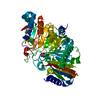
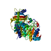
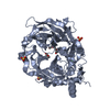
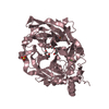
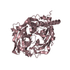
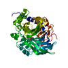
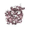
| Entry | Database: PDB / ID: 1f0i | ||||||
|---|---|---|---|---|---|---|---|
| Title | THE FIRST CRYSTAL STRUCTURE OF A PHOSPHOLIPASE D | ||||||
 Components Components | PHOSPHOLIPASE D | ||||||
 Keywords Keywords | HYDROLASE / Phospholipase D / alpha-beta-alpha-beta-alpha structure | ||||||
| Function / homology |  Function and homology information Function and homology informationphosphatidyltransferase activity / cardiolipin biosynthetic process / N-acylphosphatidylethanolamine-specific phospholipase D activity / phospholipase D / phospholipase D activity / catalytic activitySimilarity search - Function | ||||||
| Biological species |  Streptomyces sp. (bacteria) Streptomyces sp. (bacteria) | ||||||
| Method | X-RAY DIFFRACTION / SYNCHROTRON / Resolution: 1.4 Å | ||||||
 Authors Authors | Leiros, I. / Secundo, F. / Zambonelli, C. / Servi, S. / Hough, E. | ||||||
 Citation Citation | Journal: Structure Fold.Des. / Year: 2000 Title: The first crystal structure of a phospholipase D. Authors: Leiros, I. / Secundo, F. / Zambonelli, C. / Servi, S. / Hough, E. | ||||||
| History |
|
- Structure visualization
| Structure viewer | Molecule: MolmilJmol/JSmol |
|---|
- Downloads & links
-Download
| PDBx/mmCIF format | 1f0i.cif.gz | 117.3 KB | Display | PDBx/mmCIF format |
|---|---|---|---|---|
| PDB format | pdb1f0i.ent.gz | 94 KB | Display | PDB format |
| PDBx/mmJSON format | 1f0i.json.gz | Tree view | PDBx/mmJSON format | |
| Others |  Other downloads Other downloads |
-Validation report
| Arichive directory | https://data.pdbj.org/pub/pdb/validation_reports/f0/1f0iftp://data.pdbj.org/pub/pdb/validation_reports/f0/1f0i | HTTPS FTP |
|---|
-Related structure data
| Similar structure data |
|---|


-Links
 PDBj
PDBj- Assembly
| Deposited unit | 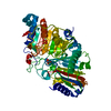
| ||||||||
|---|---|---|---|---|---|---|---|---|---|
| 1 |
| ||||||||
| Unit cell |
|
-Components
| #1: Protein | Mass: 53722.180 Da / Num. of mol.: 1 / Source method: isolated from a natural source / Source: (natural) Streptomyces sp. (bacteria) / Strain: PMFReferences: UniProt: Q93HV1, UniProt: P84147*PLUS, phospholipase D | ||
|---|---|---|---|
| #2: Chemical | Phosphate  Mass: 94.971 Da / Num. of mol.: 2 / Source method: obtained synthetically / Formula: PO4 Mass: 94.971 Da / Num. of mol.: 2 / Source method: obtained synthetically / Formula: PO4#3: Water | ChemComp-HOH / | Water Mass: 18.015 Da / Num. of mol.: 692 / Source method: isolated from a natural source / Formula: H2O Mass: 18.015 Da / Num. of mol.: 692 / Source method: isolated from a natural source / Formula: H2O |
-Experimental details
-Experiment
| Experiment | Method: X-RAY DIFFRACTION / Number of used crystals: 1 |
|---|
- Sample preparation
| Crystal | Density Matthews: 2.1 Å3/Da / Density % sol: 41.39 % | ||||||||||||||||||||
|---|---|---|---|---|---|---|---|---|---|---|---|---|---|---|---|---|---|---|---|---|---|
| Crystal grow | Temperature: 298 K / Method: vapor diffusion, sitting drop / pH: 5.4 Details: 0.2 M NH4Ac, 0.1 M Citrate/Phosphate buffer, 27.5% PEG 4000, pH 5.4, VAPOR DIFFUSION, SITTING DROP, temperature 298K | ||||||||||||||||||||
| Crystal grow | *PLUS | ||||||||||||||||||||
| Components of the solutions | *PLUS
|
-Data collection
| Diffraction | Mean temperature: 100 K |
|---|---|
| Diffraction source | Source: SYNCHROTRON / Site: ESRF  / Beamline: BM1A / Wavelength: 0.873 / Beamline: BM1A / Wavelength: 0.873 |
| Detector | Type: MARRESEARCH / Detector: IMAGE PLATE / Date: Sep 15, 1997 |
| Radiation | Protocol: SINGLE WAVELENGTH / Monochromatic (M) / Laue (L): M / Scattering type: x-ray |
| Radiation wavelength | Wavelength: 0.873 Å / Relative weight: 1 |
| Reflection | Resolution: 1.4→19.641 Å / Num. all: 86896 / Num. obs: 86896 / % possible obs: 99.2 % / Observed criterion σ(I): 3 / Redundancy: 3 % / Biso Wilson estimate: 11.652 Å2 / Rmerge(I) obs: 0.058 / Net I/σ(I): 7.4 |
| Reflection shell | Resolution: 1.4→1.48 Å / Redundancy: 3 % / Rmerge(I) obs: 0.204 / Num. unique all: 12587 / % possible all: 99.2 |
| Reflection | *PLUS Num. measured all: 261275 |
| Reflection shell | *PLUS % possible obs: 99.2 % / Mean I/σ(I) obs: 3.4 |
- Processing
| Software |
| |||||||||||||||||||||||||
|---|---|---|---|---|---|---|---|---|---|---|---|---|---|---|---|---|---|---|---|---|---|---|---|---|---|---|
| Refinement | Resolution: 1.4→10 Å / Cross valid method: THROUGHOUT / σ(F): 1 / σ(I): 1 / Stereochemistry target values: Engh & Huber
| |||||||||||||||||||||||||
| Refinement step | Cycle: LAST / Resolution: 1.4→10 Å
| |||||||||||||||||||||||||
| Refine LS restraints |
| |||||||||||||||||||||||||
| Software | *PLUS Name: SHELXL-97 / Classification: refinement | |||||||||||||||||||||||||
| Refinement | *PLUS Highest resolution: 1.4 Å / Lowest resolution: 10 Å / σ(F): 1 / % reflection Rfree: 5 % / Rfactor all: 0.13 / Rfactor obs: 0.1297 / Rfactor Rfree: 0.1823 | |||||||||||||||||||||||||
| Solvent computation | *PLUS | |||||||||||||||||||||||||
| Displacement parameters | *PLUS | |||||||||||||||||||||||||
| Refine LS restraints | *PLUS
|
