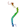
#1: Journal: J.Biol.Chem. / Year: 1998 Title: Solution Structure of a Syndecan-4 Cytoplasmic Domain and Its Interaction with Phosphatidylinositol 4,5-Bisphosphate
SYNDECAN-4 / AMPHIGLYCAN / RYUDOCAN CORE PROTEIN / SYND4
Mass: 3314.850 Da / Num. of mol.: 2 / Fragment: WHOLE CYTOPLASMIC DOMAIN / Source method: obtained synthetically Details: This peptide was chemically synthesized. The sequence of this petide occurs naturally in humans (Homo Sapiens). References: UniProt: P31431
-
Experimental details
-
Experiment
Experiment
Method: SOLUTION NMR
NMR experiment
Conditions-ID
Experiment-ID
Solution-ID
Type
1
1
1
2D NOESY
2
2
2
DQF-COSY
NMR details
Text: This structure was determined using standard 2D homonuclear techniques.
Type: Bruker DRX / Manufacturer: Bruker / Model: DRX / Field strength: 500 MHz
-
Processing
NMR software
Name
Version
Developer
Classification
XwinNMR
2.5
Bruker
collection
XwinNMR
2.5
Bruker
processing
Sparky
3.6
James, T.
dataanalysis
X-PLOR
3.851
Brunger, A.T
refinement
Refinement
Method: simulated annealing / Software ordinal: 1 Details: The structures are based on a total of 306 restraints, 246 are intramonomer NOE-derived distance constraints, 48 intermonomer NOE-derived distance restraints and 12 dihedral angle restraints.
NMR representative
Selection criteria: minimized average structure
NMR ensemble
Conformer selection criteria: structures with the lowest energy Conformers calculated total number: 90 / Conformers submitted total number: 16
+
About Yorodumi
-
News
-
Feb 9, 2022. New format data for meta-information of EMDB entries
New format data for meta-information of EMDB entries
Version 3 of the EMDB header file is now the official format.
The previous official version 1.9 will be removed from the archive.
In the structure databanks used in Yorodumi, some data are registered as the other names, "COVID-19 virus" and "2019-nCoV". Here are the details of the virus and the list of structure data.
Jan 31, 2019. EMDB accession codes are about to change! (news from PDBe EMDB page)
EMDB accession codes are about to change! (news from PDBe EMDB page)
The allocation of 4 digits for EMDB accession codes will soon come to an end. Whilst these codes will remain in use, new EMDB accession codes will include an additional digit and will expand incrementally as the available range of codes is exhausted. The current 4-digit format prefixed with “EMD-” (i.e. EMD-XXXX) will advance to a 5-digit format (i.e. EMD-XXXXX), and so on. It is currently estimated that the 4-digit codes will be depleted around Spring 2019, at which point the 5-digit format will come into force.
The EM Navigator/Yorodumi systems omit the EMD- prefix.
Related info.:Q: What is EMD? / ID/Accession-code notation in Yorodumi/EM Navigator
Yorodumi is a browser for structure data from EMDB, PDB, SASBDB, etc.
This page is also the successor to EM Navigator detail page, and also detail information page/front-end page for Omokage search.
The word "yorodu" (or yorozu) is an old Japanese word meaning "ten thousand". "mi" (miru) is to see.
Related info.:EMDB / PDB / SASBDB / Comparison of 3 databanks / Yorodumi Search / Aug 31, 2016. New EM Navigator & Yorodumi / Yorodumi Papers / Jmol/JSmol / Function and homology information / Changes in new EM Navigator and Yorodumi
 Movie
Movie Controller


 Open data
 Basic information
 Components
 Keywords
 Function and homology information
 Authors
 Citation
 Structure visualization
 Downloads & links
 Other downloads

 PDBj
PDBj


 Assembly
 Sample preparation
 Processing
 X-PLOR