 Movie
Movie Controller

[English] 日本語
 Yorodumi
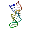
- PDB-1d31: THE THREE-DIMENSIONAL STRUCTURES OF BULGE-CONTAINING DNA FRAGMENTS -
+ Open data

- Basic information
| Entry | Database: PDB / ID: 1d31 | ||||||||||||||||||
|---|---|---|---|---|---|---|---|---|---|---|---|---|---|---|---|---|---|---|---|
| Title | THE THREE-DIMENSIONAL STRUCTURES OF BULGE-CONTAINING DNA FRAGMENTS | ||||||||||||||||||
 Components Components | DNA (5'-D(* Keywords KeywordsDNA / B-DNA / DOUBLE HELIX / BULGES / FLIPPED-OUT BASE | Function / homology | DNA / DNA (> 10) |  Function and homology information Function and homology informationMethod |  X-RAY DIFFRACTION / Resolution: 2.6 Å X-RAY DIFFRACTION / Resolution: 2.6 Å  Authors AuthorsJoshua-Tor, L. / Frolow, F. / Appella, E. / Hope, H. / Rabinovich, D. / Sussman, J.L. |  CitationJournal: J.Mol.Biol. / Year: 1992 CitationJournal: J.Mol.Biol. / Year: 1992Title: Three-dimensional structures of bulge-containing DNA fragments. Authors: Joshua-Tor, L. / Frolow, F. / Appella, E. / Hope, H. / Rabinovich, D. / Sussman, J.L. #1: Journal: Nature / Year: 1988Title: The Three-Dimensional Structure of a DNA Duplex Containing Looped-out Bases Authors: Joshua-Tor, L. / Rabinovich, D. / Hope, H. / Frolow, F. / Appella, E. / Sussman, J.L. #2: Journal: J.Mol.Biol. / Year: 1986Title: Crystallization of a DNA Tridecamer d(CGCAGAATTCGCG) Authors: Saper, M.A. / Eldar, H. / Mizuuchi, K. / Nickol, J. / Appella, E. / Sussman, J.L. History |
|
- Structure visualization
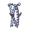
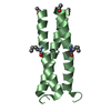
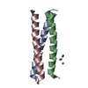
| Structure viewer | Molecule: MolmilJmol/JSmol |
|---|
- Downloads & links
-Download
| PDBx/mmCIF format | 1d31.cif.gz | 23.6 KB | Display | PDBx/mmCIF format |
|---|---|---|---|---|
| PDB format | pdb1d31.ent.gz | 15.3 KB | Display | PDB format |
| PDBx/mmJSON format | 1d31.json.gz | Tree view | PDBx/mmJSON format | |
| Others |  Other downloads Other downloads |
-Validation report
| Arichive directory | https://data.pdbj.org/pub/pdb/validation_reports/d3/1d31ftp://data.pdbj.org/pub/pdb/validation_reports/d3/1d31 | HTTPS FTP |
|---|
-Related structure data
| Similar structure data |
|---|


-Links
 PDBj
PDBj


- Assembly
| Deposited unit | 
| ||||||||
|---|---|---|---|---|---|---|---|---|---|
| 1 |
| ||||||||
| Unit cell |
|
-Components
| #1: DNA chain | Mass: 3976.599 Da / Num. of mol.: 2 / Source method: obtained synthetically #2: Water | ChemComp-HOH / |  Mass: 18.015 Da / Num. of mol.: 23 / Source method: isolated from a natural source / Formula: H2O Mass: 18.015 Da / Num. of mol.: 23 / Source method: isolated from a natural source / Formula: H2O |
|---|
-Experimental details
-Experiment
| Experiment | Method: X-RAY DIFFRACTION |
|---|
- Sample preparation
| Crystal | Density Matthews: 2.62 Å3/Da / Density % sol: 53.12 % | ||||||||||||||||||||||||||||||||||||
|---|---|---|---|---|---|---|---|---|---|---|---|---|---|---|---|---|---|---|---|---|---|---|---|---|---|---|---|---|---|---|---|---|---|---|---|---|---|
| Crystal grow | Temperature: 277 K / Method: vapor diffusion, sitting drop / pH: 7 Details: pH 7.00, VAPOR DIFFUSION, SITTING DROP, temperature 277.00K | ||||||||||||||||||||||||||||||||||||
| Components of the solutions |
|
-Data collection
| Diffraction | Mean temperature: 120 K |
|---|---|
| Diffraction source | Source: ROTATING ANODE / Type: RIGAKU / Wavelength: 1.5418 |
| Detector | Type: RIGAKU AFC-5R / Detector: DIFFRACTOMETER |
| Radiation | Scattering type: x-ray |
| Radiation wavelength | Wavelength: 1.5418 Å / Relative weight: 1 |
| Reflection | Highest resolution: 2 Å / Num. all: 6792 / Num. obs: 5530 |
| Reflection | *PLUS Highest resolution: 2 Å / Rmerge(I) obs: 0.048 |
- Processing
| Software |
| ||||||||||||
|---|---|---|---|---|---|---|---|---|---|---|---|---|---|
| Refinement | Resolution: 2.6→10 Å / σ(F): 3 /
| ||||||||||||
| Refine Biso |
| ||||||||||||
| Refinement step | Cycle: LAST / Resolution: 2.6→10 Å
| ||||||||||||
| Refinement | *PLUS Highest resolution: 2.6 Å / Lowest resolution: 10 Å / σ(F): 3 / Rfactor obs: 0.186 / Num. reflection obs: 1640 | ||||||||||||
| Solvent computation | *PLUS | ||||||||||||
| Displacement parameters | *PLUS |
