 Movie
Movie Controller

[English] 日本語
 Yorodumi
- PDB-1brp: CRYSTAL STRUCTURE OF THE TRIGONAL FORM OF HUMAN PLASMA RETINOL-BI... -
+ Open data

- Basic information
| Entry | Database: PDB / ID: 1brp | ||||||
|---|---|---|---|---|---|---|---|
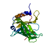
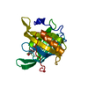
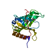
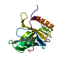
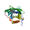
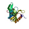
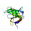
| Title | CRYSTAL STRUCTURE OF THE TRIGONAL FORM OF HUMAN PLASMA RETINOL-BINDING PROTEIN AT 2.5 ANGSTROMS RESOLUTION | ||||||
 Components Components | RETINOL BINDING PROTEIN | ||||||
 Keywords Keywords | RETINOL TRANSPORT | ||||||
| Function / homology |  Function and homology information Function and homology informationRetinoid metabolism disease events / urinary bladder development / vitamin A import into cell / embryonic retina morphogenesis in camera-type eye / retinol transport / female genitalia morphogenesis / retinol transmembrane transporter activity / embryonic organ morphogenesis / maintenance of gastrointestinal epithelium / embryonic skeletal system development ...Retinoid metabolism disease events / urinary bladder development / vitamin A import into cell / embryonic retina morphogenesis in camera-type eye / retinol transport / female genitalia morphogenesis / retinol transmembrane transporter activity / embryonic organ morphogenesis / maintenance of gastrointestinal epithelium / embryonic skeletal system development / negative regulation of cardiac muscle cell proliferation / detection of light stimulus involved in visual perception / retinal metabolic process / eye development / retinal binding / heart trabecula formation / molecular carrier activity / retinol metabolic process / retinol binding / cardiac muscle tissue development / positive regulation of immunoglobulin production / Defective visual phototransduction due to STRA6 loss of function / response to muscle activity / The canonical retinoid cycle in rods (twilight vision) / uterus development / vagina development / phototransduction, visible light / response to retinoic acid / Retinoid metabolism and transport / lung development / gluconeogenesis / positive regulation of insulin secretion / response to insulin / male gonad development / glucose homeostasis / heart development / spermatogenesis / response to ethanol / response to xenobiotic stimulus / protein-containing complex binding / protein-containing complex / extracellular space / extracellular exosome / extracellular region Similarity search - Function | ||||||
| Biological species |  Homo sapiens (human) Homo sapiens (human) | ||||||
| Method |  X-RAY DIFFRACTION / Resolution: 2.5 Å X-RAY DIFFRACTION / Resolution: 2.5 Å | ||||||
 Authors Authors | Zanotti, G. / Monaco, H.L. | ||||||
 Citation Citation | Journal: J.Mol.Biol. / Year: 1993 Title: Crystal structure of the trigonal form of human plasma retinol-binding protein at 2.5 A resolution. Authors: Zanotti, G. / Ottonello, S. / Berni, R. / Monaco, H.L. #1: Journal: J.Mol.Biol. / Year: 1984Title: Crystallization of Human Plasma Apo-Retinol-Binding Protein Authors: Monaco, H.L. / Zanotti, G. / Ottonello, S. / Berni, R. #2: Journal: J.Mol.Biol. / Year: 1983Title: Crystallization and Preliminary X-Ray Data of Human Plasma Retinol-Binding Protein Authors: Ottonello, S. / Maraini, G. / Mammi, M. / Monaco, H.L. / Spadon, P. / Zanotti, G. | ||||||
| History |
| ||||||
| Remark 700 | SHEET THE SHEET PRESENTED AS *S1* ON SHEET RECORDS BELOW IS ACTUALLY AN EIGHT-STRANDED BETA-BARREL. ...SHEET THE SHEET PRESENTED AS *S1* ON SHEET RECORDS BELOW IS ACTUALLY AN EIGHT-STRANDED BETA-BARREL. THIS IS REPRESENTED BY A NINE-STRANDED SHEET IN WHICH THE FIRST AND LAST STRANDS ARE IDENTICAL. |
- Structure visualization
| Structure viewer | Molecule: MolmilJmol/JSmol |
|---|
- Downloads & links
-Download
| PDBx/mmCIF format | 1brp.cif.gz | 49.7 KB | Display | PDBx/mmCIF format |
|---|---|---|---|---|
| PDB format | pdb1brp.ent.gz | 35.4 KB | Display | PDB format |
| PDBx/mmJSON format | 1brp.json.gz | Tree view | PDBx/mmJSON format | |
| Others |  Other downloads Other downloads |
-Validation report
| Arichive directory | https://data.pdbj.org/pub/pdb/validation_reports/br/1brpftp://data.pdbj.org/pub/pdb/validation_reports/br/1brp | HTTPS FTP |
|---|
-Related structure data


-Links
 PDBj
PDBj


- Assembly
| Deposited unit | 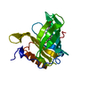
| ||||||||
|---|---|---|---|---|---|---|---|---|---|
| 1 |
| ||||||||
| Unit cell |
| ||||||||
| Atom site foot note | 1: DISORDERED AREAS: RESIDUES 1 - 3, 64 - 67, AND C TERMINAL PORTION (RESIDUES 175 - 182) CANNOT BE SEEN IN THE MAP. |
-Components
| #1: Protein | Mass: 20984.445 Da / Num. of mol.: 1 Source method: isolated from a genetically manipulated source Source: (gene. exp.) Homo sapiens (human) / References: UniProt: P02753 |
|---|---|
| #2: Chemical | ChemComp-RTL /   Mass: 286.452 Da / Num. of mol.: 1 / Source method: obtained synthetically / Formula: C20H30O Mass: 286.452 Da / Num. of mol.: 1 / Source method: obtained synthetically / Formula: C20H30O |
| #3: Water | ChemComp-HOH /  Mass: 18.015 Da / Num. of mol.: 54 / Source method: isolated from a natural source / Formula: H2O Mass: 18.015 Da / Num. of mol.: 54 / Source method: isolated from a natural source / Formula: H2O |
| Has protein modification | Y |
-Experimental details
-Experiment
| Experiment | Method: X-RAY DIFFRACTION |
|---|
- Sample preparation
| Crystal | Density Matthews: 3.69 Å3/Da / Density % sol: 66.66 % | ||||||||||||||||||||||||||||||||||||||||||
|---|---|---|---|---|---|---|---|---|---|---|---|---|---|---|---|---|---|---|---|---|---|---|---|---|---|---|---|---|---|---|---|---|---|---|---|---|---|---|---|---|---|---|---|
| Crystal grow | *PLUS Temperature: 4 ℃ / pH: 6.8 / Method: microdialysis / Details: referred to J.Mol.Biol. 163.679-681 1983 | ||||||||||||||||||||||||||||||||||||||||||
| Components of the solutions | *PLUS
|
-Data collection
| Radiation | Scattering type: x-ray |
|---|---|
| Radiation wavelength | Relative weight: 1 |
| Reflection | *PLUS Highest resolution: 2.5 Å / Lowest resolution: 9999 Å / Num. obs: 9828 / Num. measured all: 29524 / Rmerge(I) obs: 0.096 |
| Reflection shell | *PLUS Highest resolution: 2.5 Å / Lowest resolution: 2.7 Å / % possible obs: 76 % / Num. possible: 76 / Num. measured obs: 1566 |
- Processing
| Software |
| ||||||||||||||||||||||||||||||||||||||||||||||||||||||||||||
|---|---|---|---|---|---|---|---|---|---|---|---|---|---|---|---|---|---|---|---|---|---|---|---|---|---|---|---|---|---|---|---|---|---|---|---|---|---|---|---|---|---|---|---|---|---|---|---|---|---|---|---|---|---|---|---|---|---|---|---|---|---|
| Refinement | Highest resolution: 2.5 Å /
| ||||||||||||||||||||||||||||||||||||||||||||||||||||||||||||
| Refinement step | Cycle: LAST / Highest resolution: 2.5 Å
| ||||||||||||||||||||||||||||||||||||||||||||||||||||||||||||
| Refine LS restraints |
| ||||||||||||||||||||||||||||||||||||||||||||||||||||||||||||
| Software | *PLUS Name: X-PLOR / Classification: refinement | ||||||||||||||||||||||||||||||||||||||||||||||||||||||||||||
| Refinement | *PLUS Highest resolution: 2.5 Å / Lowest resolution: 9 Å / Num. reflection obs: 9652 / Rfactor obs: 0.176 | ||||||||||||||||||||||||||||||||||||||||||||||||||||||||||||
| Solvent computation | *PLUS | ||||||||||||||||||||||||||||||||||||||||||||||||||||||||||||
| Displacement parameters | *PLUS | ||||||||||||||||||||||||||||||||||||||||||||||||||||||||||||
| Refine LS restraints | *PLUS
|
