 Movie
Movie Controller


+ Open data

- Basic information
| Entry | Database: PDB / ID: 1b5o | ||||||
|---|---|---|---|---|---|---|---|
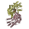
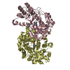
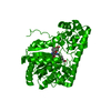
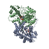
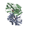
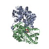
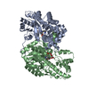
| Title | THERMUS THERMOPHILUS ASPARTATE AMINOTRANSFERASE SINGLE MUTANT 1 | ||||||
 Components Components | PROTEIN (ASPARTATE AMINOTRANSFERASE) | ||||||
 Keywords Keywords |  TRANSFERASE / AMINOTRANSFERASE / PYRIDOXAL ENZYME TRANSFERASE / AMINOTRANSFERASE / PYRIDOXAL ENZYME | ||||||
| Function / homology |  Function and homology informationaspartate-prephenate aminotransferase / aspartate-prephenate aminotransferase activity / aspartate transaminase / L-aspartate:2-oxoglutarate aminotransferase activity / biosynthetic process / pyridoxal phosphate binding / cytoplasm Function and homology informationaspartate-prephenate aminotransferase / aspartate-prephenate aminotransferase activity / aspartate transaminase / L-aspartate:2-oxoglutarate aminotransferase activity / biosynthetic process / pyridoxal phosphate binding / cytoplasmSimilarity search - Function | ||||||
| Biological species |   Thermus thermophilus (bacteria) Thermus thermophilus (bacteria) | ||||||
| Method | X-RAY DIFFRACTION / SYNCHROTRON / MOLECULAR REPLACEMENT / Resolution: 2.2 Å | ||||||
 Authors Authors | Ura, H. / Nakai, T. / Kawaguchi, S.I. / Miyahara, I. / Hirotsu, K. / Kuramitsu, S. | ||||||
 Citation Citation | Journal: J.BIOCHEM.(TOKYO) / Year: 2001 Title: Substrate recognition mechanism of thermophilic dual-substrate enzyme Authors: Ura, H. / Nakai, T. / Kawaguchi, S.I. / Miyahara, I. / Hirotsu, K. / Kuramitsu, S. | ||||||
| History |
|
- Structure visualization
| Structure viewer | Molecule: MolmilJmol/JSmol |
|---|
- Downloads & links
-Download
| PDBx/mmCIF format | 1b5o.cif.gz | 159.8 KB | Display | PDBx/mmCIF format |
|---|---|---|---|---|
| PDB format | pdb1b5o.ent.gz | 126.8 KB | Display | PDB format |
| PDBx/mmJSON format | 1b5o.json.gz | Tree view | PDBx/mmJSON format | |
| Others |  Other downloads Other downloads |
-Validation report
| Arichive directory | https://data.pdbj.org/pub/pdb/validation_reports/b5/1b5oftp://data.pdbj.org/pub/pdb/validation_reports/b5/1b5o | HTTPS FTP |
|---|
-Related structure data
| Related structure data |  1b5pC  1gc3C  1gc4C  1gckC  5bj3C  5bj4C 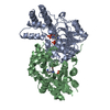 1bjwS S: Starting model for refinement C: citing same article ( |
|---|---|
| Similar structure data |


-Links
 PDBj
PDBj- Assembly
| Deposited unit | 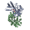
| ||||||||
|---|---|---|---|---|---|---|---|---|---|
| 1 |
| ||||||||
| Unit cell |
|
-Components
| #1: Protein | Mass: 42060.832 Da / Num. of mol.: 2 / Mutation: K101S Source method: isolated from a genetically manipulated source Source: (gene. exp.) Thermus thermophilus (bacteria) / Strain: HB8 / Production host: Escherichia coli (E. coli) / Strain (production host): AB1157 / References: UniProt: Q56232, aspartate transaminase#2: Chemical | Phosphate  Mass: 94.971 Da / Num. of mol.: 2 / Source method: obtained synthetically / Formula: PO4 Mass: 94.971 Da / Num. of mol.: 2 / Source method: obtained synthetically / Formula: PO4#3: Chemical | Pyridoxal phosphate  Mass: 247.142 Da / Num. of mol.: 2 / Source method: obtained synthetically / Formula: C8H10NO6P Mass: 247.142 Da / Num. of mol.: 2 / Source method: obtained synthetically / Formula: C8H10NO6P#4: Water | ChemComp-HOH / | Water Mass: 18.015 Da / Num. of mol.: 277 / Source method: isolated from a natural source / Formula: H2O Mass: 18.015 Da / Num. of mol.: 277 / Source method: isolated from a natural source / Formula: H2O |
|---|
-Experimental details
-Experiment
| Experiment | Method: X-RAY DIFFRACTION / Number of used crystals: 1 |
|---|
- Sample preparation
| Crystal | Density Matthews: 2.57 Å3/Da / Density % sol: 52 % | |||||||||||||||||||||||||||||||||||
|---|---|---|---|---|---|---|---|---|---|---|---|---|---|---|---|---|---|---|---|---|---|---|---|---|---|---|---|---|---|---|---|---|---|---|---|---|
| Crystal grow | pH: 4.3 / Details: CRYSTALLIZED FROM 300MM AMMONIUM PHOSPHATE, PH 4.3 | |||||||||||||||||||||||||||||||||||
| Crystal grow | *PLUS pH: 8 / Method: vapor diffusion, hanging drop | |||||||||||||||||||||||||||||||||||
| Components of the solutions | *PLUS
|
-Data collection
| Diffraction | Mean temperature: 287 K |
|---|---|
| Diffraction source | Source: SYNCHROTRON / Site: Photon Factory  / Beamline: BL-6A / Wavelength: 1 / Beamline: BL-6A / Wavelength: 1 |
| Detector | Type: RIGAKU / Detector: IMAGE PLATE / Date: Jun 15, 1998 / Details: MIRROR |
| Radiation | Protocol: SINGLE WAVELENGTH / Monochromatic (M) / Laue (L): M / Scattering type: x-ray |
| Radiation wavelength | Wavelength: 1 Å / Relative weight: 1 |
| Reflection | Resolution: 2.2→50 Å / Num. obs: 40675 / % possible obs: 88.5 % / Observed criterion σ(I): 0 / Redundancy: 2.2 % / Biso Wilson estimate: 20.6 Å2 / Rmerge(I) obs: 0.067 / Net I/σ(I): 16 |
| Reflection shell | Resolution: 2.2→2.28 Å / Redundancy: 1.8 % / Rmerge(I) obs: 0.133 / Mean I/σ(I) obs: 5.3 / % possible all: 74.3 |
| Reflection | *PLUS Num. measured all: 89485 |
- Processing
| Software |
| ||||||||||||||||||||||||||||||||||||||||||||||||||||||||||||||||||||||||||||||||
|---|---|---|---|---|---|---|---|---|---|---|---|---|---|---|---|---|---|---|---|---|---|---|---|---|---|---|---|---|---|---|---|---|---|---|---|---|---|---|---|---|---|---|---|---|---|---|---|---|---|---|---|---|---|---|---|---|---|---|---|---|---|---|---|---|---|---|---|---|---|---|---|---|---|---|---|---|---|---|---|---|---|
| Refinement | Method to determine structure: MOLECULAR REPLACEMENT Starting model: PDB ENTRY 1BJW Resolution: 2.2→8 Å / Rfactor Rfree error: 0.0038 / Data cutoff high absF: 1000000 / Data cutoff low absF: 0.001 / Isotropic thermal model: RESTRAINED / Cross valid method: THROUGHOUT / σ(F): 2
| ||||||||||||||||||||||||||||||||||||||||||||||||||||||||||||||||||||||||||||||||
| Displacement parameters | Biso mean: 19.6 Å2 | ||||||||||||||||||||||||||||||||||||||||||||||||||||||||||||||||||||||||||||||||
| Refinement step | Cycle: LAST / Resolution: 2.2→8 Å
| ||||||||||||||||||||||||||||||||||||||||||||||||||||||||||||||||||||||||||||||||
| Refine LS restraints |
| ||||||||||||||||||||||||||||||||||||||||||||||||||||||||||||||||||||||||||||||||
| LS refinement shell | Resolution: 2.2→2.3 Å / Rfactor Rfree error: 0.012 / Total num. of bins used: 8
| ||||||||||||||||||||||||||||||||||||||||||||||||||||||||||||||||||||||||||||||||
| Xplor file |
| ||||||||||||||||||||||||||||||||||||||||||||||||||||||||||||||||||||||||||||||||
| Refinement | *PLUS % reflection Rfree: 10 % | ||||||||||||||||||||||||||||||||||||||||||||||||||||||||||||||||||||||||||||||||
| Solvent computation | *PLUS | ||||||||||||||||||||||||||||||||||||||||||||||||||||||||||||||||||||||||||||||||
| Displacement parameters | *PLUS | ||||||||||||||||||||||||||||||||||||||||||||||||||||||||||||||||||||||||||||||||
| Refine LS restraints | *PLUS
|
