 Movie
Movie Controller


+ Open data

- Basic information
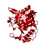
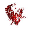
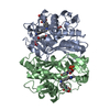
| Entry | Database: PDB / ID: 1aob | ||||||
|---|---|---|---|---|---|---|---|
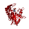
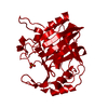
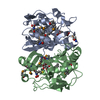
| Title | E. COLI THYMIDYLATE SYNTHASE COMPLEXED WITH DDURD | ||||||
 Components Components | THYMIDYLATE SYNTHASE | ||||||
 Keywords Keywords | METHYLTRANSFERASE / TRANSFERASE (METHYLTRANSFERASE) / SUBSTRATE MODULES | ||||||
| Function / homology |  Function and homology information Function and homology informationthymidylate synthase / thymidylate synthase activity / dTMP biosynthetic process / dTTP biosynthetic process / response to radiation / regulation of translation / methylation / magnesium ion binding / protein homodimerization activity / RNA binding / cytosol Similarity search - Function | ||||||
| Biological species |  | ||||||
| Method |  X-RAY DIFFRACTION / DIFFERENCE FOURIER / Resolution: 2.1 Å X-RAY DIFFRACTION / DIFFERENCE FOURIER / Resolution: 2.1 Å | ||||||
 Authors Authors | Stout, T.J. / Sage, C.R. / Stroud, R.M. | ||||||
 Citation Citation | Journal: Structure / Year: 1998 Title: The additivity of substrate fragments in enzyme-ligand binding. Authors: Stout, T.J. / Sage, C.R. / Stroud, R.M. | ||||||
| History |
|
- Structure visualization
| Structure viewer | Molecule: MolmilJmol/JSmol |
|---|
- Downloads & links
-Download
| PDBx/mmCIF format | 1aob.cif.gz | 62.5 KB | Display | PDBx/mmCIF format |
|---|---|---|---|---|
| PDB format | pdb1aob.ent.gz | 49.1 KB | Display | PDB format |
| PDBx/mmJSON format | 1aob.json.gz | Tree view | PDBx/mmJSON format | |
| Others |  Other downloads Other downloads |
-Validation report
| Arichive directory | https://data.pdbj.org/pub/pdb/validation_reports/ao/1aobftp://data.pdbj.org/pub/pdb/validation_reports/ao/1aob | HTTPS FTP |
|---|
-Related structure data
| Related structure data |  1an5C  1bduC  1bidC  1dduC  1tduC  1tjsSC 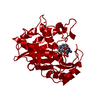 1trgC S: Starting model for refinement C: citing same article ( |
|---|---|
| Similar structure data |


-Links
 PDBj
PDBj- Assembly
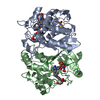
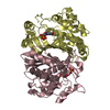
| Deposited unit | 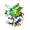
| ||||||||
|---|---|---|---|---|---|---|---|---|---|
| 1 | 
| ||||||||
| Unit cell |
|
-Components
| #1: Protein | Mass: 30515.654 Da / Num. of mol.: 1 Source method: isolated from a genetically manipulated source Source: (gene. exp.) |
|---|---|
| #2: Chemical | ChemComp-PO4 /   Mass: 94.971 Da / Num. of mol.: 1 / Source method: obtained synthetically / Formula: PO4 Mass: 94.971 Da / Num. of mol.: 1 / Source method: obtained synthetically / Formula: PO4 |
| #3: Chemical | ChemComp-DDU /   Mass: 212.203 Da / Num. of mol.: 1 / Source method: obtained synthetically / Formula: C9H12N2O4 Mass: 212.203 Da / Num. of mol.: 1 / Source method: obtained synthetically / Formula: C9H12N2O4 |
| #4: Chemical | ChemComp-FMT /   Mass: 46.025 Da / Num. of mol.: 1 / Source method: obtained synthetically / Formula: CH2O2 Mass: 46.025 Da / Num. of mol.: 1 / Source method: obtained synthetically / Formula: CH2O2 |
-Experimental details
-Experiment
| Experiment | Method: X-RAY DIFFRACTION / Number of used crystals: 1 |
|---|
- Sample preparation
| Crystal | Density Matthews: 3.3 Å3/Da / Density % sol: 58.1 % | ||||||||||||||||||||||||||||||||||||||||
|---|---|---|---|---|---|---|---|---|---|---|---|---|---|---|---|---|---|---|---|---|---|---|---|---|---|---|---|---|---|---|---|---|---|---|---|---|---|---|---|---|---|
| Crystal grow | Method: vapor diffusion, hanging drop / pH: 7.8 Details: CRYSTALLIZATION EXPERIMENTS WERE CONDUCTED IN HANGING DROPS CONTAINING 4.2 MG/ML E. COLI TS, 0.38 MM DDURD, 3.8 MM DTT, AND 1.2 M (NH4)2SO4, AT PH 7.8 (20 MM KPO4) SUSPENDED OVER A WELL ...Details: CRYSTALLIZATION EXPERIMENTS WERE CONDUCTED IN HANGING DROPS CONTAINING 4.2 MG/ML E. COLI TS, 0.38 MM DDURD, 3.8 MM DTT, AND 1.2 M (NH4)2SO4, AT PH 7.8 (20 MM KPO4) SUSPENDED OVER A WELL SOLUTION CONTAINING 2.4 M (NH4)2SO4 AND 1.0 MM DTT., vapor diffusion - hanging drop | ||||||||||||||||||||||||||||||||||||||||
| Crystal | *PLUS | ||||||||||||||||||||||||||||||||||||||||
| Crystal grow | *PLUS Method: vapor diffusion, hanging drop | ||||||||||||||||||||||||||||||||||||||||
| Components of the solutions | *PLUS
|
-Data collection
| Diffraction | Mean temperature: 298 K |
|---|---|
| Diffraction source | Source: ROTATING ANODE / Type: RIGAKU RUH2R / Wavelength: 1.5418 |
| Detector | Type: RIGAKU / Detector: IMAGE PLATE / Date: Nov 1, 1996 |
| Radiation | Monochromator: GRAPHITE(002) / Monochromatic (M) / Laue (L): M / Scattering type: x-ray |
| Radiation wavelength | Wavelength: 1.5418 Å / Relative weight: 1 |
| Reflection | Resolution: 2.1→30 Å / Num. obs: 22981 / % possible obs: 89.7 % / Observed criterion σ(I): 1 / Redundancy: 12.4 % / Biso Wilson estimate: 9.7 Å2 / Rmerge(I) obs: 0.098 / Rsym value: 0.098 / Net I/σ(I): 11.9 |
| Reflection shell | Resolution: 2.1→2.16 Å / Redundancy: 2.4 % / Rmerge(I) obs: 0.34 / Mean I/σ(I) obs: 1.9 / Rsym value: 0.34 / % possible all: 74.5 |
| Reflection | *PLUS Num. measured all: 76509 |
- Processing
| Software |
| ||||||||||||||||||||||||||||||||||||||||||||||||||||||||||||||||||||||||||||||||
|---|---|---|---|---|---|---|---|---|---|---|---|---|---|---|---|---|---|---|---|---|---|---|---|---|---|---|---|---|---|---|---|---|---|---|---|---|---|---|---|---|---|---|---|---|---|---|---|---|---|---|---|---|---|---|---|---|---|---|---|---|---|---|---|---|---|---|---|---|---|---|---|---|---|---|---|---|---|---|---|---|---|
| Refinement | Method to determine structure: DIFFERENCE FOURIER Starting model: PDB ENTRY 1TJS Resolution: 2.1→8 Å / Rfactor Rfree error: 0.006 / Data cutoff high absF: 10000000 / Data cutoff low absF: 0.001 / Isotropic thermal model: RESTRAINED / Cross valid method: THROUGHOUT / σ(F): 1
| ||||||||||||||||||||||||||||||||||||||||||||||||||||||||||||||||||||||||||||||||
| Displacement parameters | Biso mean: 22.3 Å2 | ||||||||||||||||||||||||||||||||||||||||||||||||||||||||||||||||||||||||||||||||
| Refine analyze |
| ||||||||||||||||||||||||||||||||||||||||||||||||||||||||||||||||||||||||||||||||
| Refinement step | Cycle: LAST / Resolution: 2.1→8 Å
| ||||||||||||||||||||||||||||||||||||||||||||||||||||||||||||||||||||||||||||||||
| Refine LS restraints |
| ||||||||||||||||||||||||||||||||||||||||||||||||||||||||||||||||||||||||||||||||
| LS refinement shell | Resolution: 2.1→2.23 Å / Rfactor Rfree error: 0.022 / Total num. of bins used: 6
| ||||||||||||||||||||||||||||||||||||||||||||||||||||||||||||||||||||||||||||||||
| Xplor file |
| ||||||||||||||||||||||||||||||||||||||||||||||||||||||||||||||||||||||||||||||||
| Software | *PLUS Name: X-PLOR / Version: 3.843 / Classification: refinement | ||||||||||||||||||||||||||||||||||||||||||||||||||||||||||||||||||||||||||||||||
| Refine LS restraints | *PLUS
|
