 Movie
Movie Controller


+ Open data

- Basic information
| Entry | Database: PDB / ID: 1an4 | ||||||
|---|---|---|---|---|---|---|---|
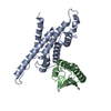
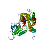
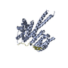
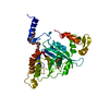
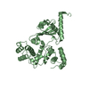
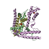
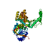
| Title | STRUCTURE AND FUNCTION OF THE B/HLH/Z DOMAIN OF USF | ||||||
 Components Components |
| ||||||
 Keywords Keywords | TRANSCRIPTION/DNA / PROTEIN-DNA COMPLEX / DOUBLE HELIX / OVERHANGING BASE / TRANSCRIPTION-DNA COMPLEX | ||||||
| Function / homology |  Function and homology information Function and homology informationcarbon catabolite regulation of transcription / regulation of transcription from RNA polymerase II promoter by glucose / late viral transcription / glucose mediated signaling pathway / positive regulation of transcription from RNA polymerase II promoter by glucose / bHLH transcription factor binding / lipid homeostasis / Regulation of MITF-M-dependent genes involved in pigmentation / negative regulation of fibrinolysis / response to UV ...carbon catabolite regulation of transcription / regulation of transcription from RNA polymerase II promoter by glucose / late viral transcription / glucose mediated signaling pathway / positive regulation of transcription from RNA polymerase II promoter by glucose / bHLH transcription factor binding / lipid homeostasis / Regulation of MITF-M-dependent genes involved in pigmentation / negative regulation of fibrinolysis / response to UV / cellular response to glucose stimulus / cellular response to insulin stimulus / glucose metabolic process / histone deacetylase binding / sequence-specific double-stranded DNA binding / glucose homeostasis / DNA-binding transcription activator activity, RNA polymerase II-specific / transcription regulator complex / Estrogen-dependent gene expression / sequence-specific DNA binding / DNA-binding transcription factor activity, RNA polymerase II-specific / response to hypoxia / RNA polymerase II cis-regulatory region sequence-specific DNA binding / protein heterodimerization activity / regulation of transcription by RNA polymerase II / protein kinase binding / chromatin / protein-containing complex binding / enzyme binding / Golgi apparatus / protein homodimerization activity / positive regulation of transcription by RNA polymerase II / nucleoplasm / identical protein binding / nucleus Similarity search - Function | ||||||
| Biological species |  Homo sapiens (human) Homo sapiens (human) | ||||||
| Method |  X-RAY DIFFRACTION / SYNCHROTRON / Resolution: 2.9 Å X-RAY DIFFRACTION / SYNCHROTRON / Resolution: 2.9 Å | ||||||
 Authors Authors | Ferre-D'Amare, A.R. / Pognonec, P. / Roeder, R.G. / Burley, S.K. | ||||||
 Citation Citation | Journal: EMBO J. / Year: 1994 Title: Structure and function of the b/HLH/Z domain of USF. Authors: Ferre-D'Amare, A.R. / Pognonec, P. / Roeder, R.G. / Burley, S.K. | ||||||
| History |
|
- Structure visualization
| Structure viewer | Molecule: MolmilJmol/JSmol |
|---|
- Downloads & links
-Download
| PDBx/mmCIF format | 1an4.cif.gz | 66 KB | Display | PDBx/mmCIF format |
|---|---|---|---|---|
| PDB format | pdb1an4.ent.gz | 44.9 KB | Display | PDB format |
| PDBx/mmJSON format | 1an4.json.gz | Tree view | PDBx/mmJSON format | |
| Others |  Other downloads Other downloads |
-Validation report
| Arichive directory | https://data.pdbj.org/pub/pdb/validation_reports/an/1an4ftp://data.pdbj.org/pub/pdb/validation_reports/an/1an4 | HTTPS FTP |
|---|
-Related structure data
| Similar structure data |
|---|


-Links
 PDBj
PDBj


- Assembly
| Deposited unit | 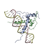
| ||||||||
|---|---|---|---|---|---|---|---|---|---|
| 1 |
| ||||||||
| Unit cell |
|
-Components
| #1: DNA chain | Mass: 6369.113 Da / Num. of mol.: 1 Source method: isolated from a genetically manipulated source |
|---|---|
| #2: DNA chain | Mass: 6520.195 Da / Num. of mol.: 1 Source method: isolated from a genetically manipulated source |
| #3: Protein | Mass: 7653.597 Da / Num. of mol.: 2 Fragment: FRAGMENT:B/HLH DNA BINDING DOMAIN MUTATION:R196M, C229S, C248S Mutation: R196M, C229S, C248S Source method: isolated from a genetically manipulated source Source: (gene. exp.) Homo sapiens (human) / Production host: Bacteria (eubacteria) / Keywords: B/HLH DNA BINDING DOMAIN / References: UniProt: P22415 |
-Experimental details
-Experiment
| Experiment | Method: X-RAY DIFFRACTION |
|---|
- Sample preparation
| Crystal | Density Matthews: 2.88 Å3/Da / Density % sol: 57.35 % | ||||||||||||||||||||||||||||||||||||||||||||||||||||||||||||||||||||||||||||||||||||||||||
|---|---|---|---|---|---|---|---|---|---|---|---|---|---|---|---|---|---|---|---|---|---|---|---|---|---|---|---|---|---|---|---|---|---|---|---|---|---|---|---|---|---|---|---|---|---|---|---|---|---|---|---|---|---|---|---|---|---|---|---|---|---|---|---|---|---|---|---|---|---|---|---|---|---|---|---|---|---|---|---|---|---|---|---|---|---|---|---|---|---|---|---|
| Crystal grow | Temperature: 277 K / Method: vapor diffusion, hanging drop / pH: 4.75 Details: pH 4.75, VAPOR DIFFUSION, HANGING DROP, temperature 277.00K | ||||||||||||||||||||||||||||||||||||||||||||||||||||||||||||||||||||||||||||||||||||||||||
| Components of the solutions |
| ||||||||||||||||||||||||||||||||||||||||||||||||||||||||||||||||||||||||||||||||||||||||||
| Crystal grow | *PLUS Temperature: 4 ℃ / pH: 7.5 | ||||||||||||||||||||||||||||||||||||||||||||||||||||||||||||||||||||||||||||||||||||||||||
| Components of the solutions | *PLUS
|
-Data collection
| Diffraction | Mean temperature: 253 K |
|---|---|
| Diffraction source | Source: SYNCHROTRON / Site: CHESS  / Beamline: F1 / Beamline: F1 |
| Detector | Detector: IMAGE PLATE / Date: Jun 1, 1993 |
| Radiation | Scattering type: x-ray |
| Radiation wavelength | Relative weight: 1 |
| Reflection | Resolution: 2.9→15 Å / Num. all: 32049 / Num. obs: 6038 / % possible obs: 77.2 % / Redundancy: 2.6 % / Rmerge(I) obs: 0.076 / Net I/σ(I): 14.4 |
| Reflection shell | Resolution: 2.9→3 Å / Redundancy: 1.5 % / % possible all: 49.3 |
| Reflection | *PLUS Highest resolution: 2.9 Å / Lowest resolution: 15 Å / % possible obs: 76.4 % / Observed criterion σ(I): 1 / Redundancy: 2.6 % / Num. measured all: 32049 |
| Reflection shell | *PLUS Highest resolution: 2.9 Å / Lowest resolution: 3 Å / % possible obs: 49.3 % / Redundancy: 1.5 % |
- Processing
| Software |
| ||||||||||||||||||||||||||||||||||||||||||||||||||||||||||||
|---|---|---|---|---|---|---|---|---|---|---|---|---|---|---|---|---|---|---|---|---|---|---|---|---|---|---|---|---|---|---|---|---|---|---|---|---|---|---|---|---|---|---|---|---|---|---|---|---|---|---|---|---|---|---|---|---|---|---|---|---|---|
| Refinement | Resolution: 2.9→6 Å / Rfactor Rwork: 0.236 / σ(F): 2 | ||||||||||||||||||||||||||||||||||||||||||||||||||||||||||||
| Refinement step | Cycle: LAST / Resolution: 2.9→6 Å
| ||||||||||||||||||||||||||||||||||||||||||||||||||||||||||||
| Refine LS restraints |
| ||||||||||||||||||||||||||||||||||||||||||||||||||||||||||||
| Software | *PLUS Name: X-PLOR / Version: 3.1 / Classification: refinement | ||||||||||||||||||||||||||||||||||||||||||||||||||||||||||||
| Refinement | *PLUS Highest resolution: 2.9 Å / Lowest resolution: 6 Å / Num. reflection obs: 5096 / σ(F): 1 | ||||||||||||||||||||||||||||||||||||||||||||||||||||||||||||
| Solvent computation | *PLUS | ||||||||||||||||||||||||||||||||||||||||||||||||||||||||||||
| Displacement parameters | *PLUS | ||||||||||||||||||||||||||||||||||||||||||||||||||||||||||||
| Refine LS restraints | *PLUS Type: x_angle_deg / Dev ideal: 3.05 |
