 Movie
Movie Controller

[English] 日本語
 Yorodumi
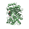
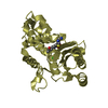
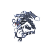
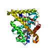
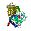
- PDB-1alq: CIRCULARLY PERMUTED BETA-LACTAMASE FROM STAPHYLOCOCCUS AUREUS PC1 -
+ Open data

- Basic information
| Entry | Database: PDB / ID: 1alq | ||||||
|---|---|---|---|---|---|---|---|
| Title | CIRCULARLY PERMUTED BETA-LACTAMASE FROM STAPHYLOCOCCUS AUREUS PC1 | ||||||
 Components Components | CP254 BETA-LACTAMASE | ||||||
 Keywords Keywords | HYDROLASE / CIRCULAR PERMUTED / ANTIBIOTIC RESISTANCE | ||||||
| Function / homology |  Function and homology information Function and homology informationbeta-lactam antibiotic catabolic process / beta-lactamase activity / beta-lactamase / response to antibiotic Similarity search - Function | ||||||
| Biological species |   Staphylococcus aureus (bacteria) Staphylococcus aureus (bacteria) | ||||||
| Method |  X-RAY DIFFRACTION / DIFFERENCE FOURIER / Resolution: 1.8 Å X-RAY DIFFRACTION / DIFFERENCE FOURIER / Resolution: 1.8 Å | ||||||
 Authors Authors | Pieper, U. / Herzberg, O. | ||||||
 Citation Citation | Journal: Biochemistry / Year: 1997 Title: Circularly permuted beta-lactamase from Staphylococcus aureus PC1. Authors: Pieper, U. / Hayakawa, K. / Li, Z. / Herzberg, O. #1: Journal: J.Mol.Biol. / Year: 1991Title: Refined Crystal Structure of Beta-Lactamase from Staphylococcus Aureus Pc1 at 2.0 A Resolution Authors: Herzberg, O. #2: Journal: Science / Year: 1987Title: Bacterial Resistance to Beta-Lactam Antibiotics: Crystal Structure of Beta-Lactamase from Staphylococcus Aureus Pc1 at 2.5 A Resolution Authors: Herzberg, O. / Moult, J. | ||||||
| History |
|
- Structure visualization
| Structure viewer | Molecule: MolmilJmol/JSmol |
|---|
- Downloads & links
-Download
| PDBx/mmCIF format | 1alq.cif.gz | 69.5 KB | Display | PDBx/mmCIF format |
|---|---|---|---|---|
| PDB format | pdb1alq.ent.gz | 50 KB | Display | PDB format |
| PDBx/mmJSON format | 1alq.json.gz | Tree view | PDBx/mmJSON format | |
| Others |  Other downloads Other downloads |
-Validation report
| Arichive directory | https://data.pdbj.org/pub/pdb/validation_reports/al/1alqftp://data.pdbj.org/pub/pdb/validation_reports/al/1alq | HTTPS FTP |
|---|
-Related structure data
| Related structure data |  3blmS S: Starting model for refinement |
|---|---|
| Similar structure data |


-Links
 PDBj
PDBj


- Assembly
| Deposited unit | 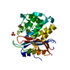
| ||||||||||||
|---|---|---|---|---|---|---|---|---|---|---|---|---|---|
| 1 |
| ||||||||||||
| Unit cell |
| ||||||||||||
| Components on special symmetry positions |
|
-Components
| #1: Protein | Mass: 29646.125 Da / Num. of mol.: 1 Mutation: CIRCULARLY PERMUTED WITH AN EIGHT RESIDUE LINKER INSERTED TO JOIN THE ORIGINAL N- AND C-TERMINI, AND A NEW N-TERMINUS AT RESIDUE 254 Source method: isolated from a genetically manipulated source Source: (gene. exp.) Staphylococcus aureus (bacteria) / Cell line: 293 / Production host: |
|---|---|
| #2: Chemical | ChemComp-SO4 /   Mass: 96.063 Da / Num. of mol.: 1 / Source method: obtained synthetically / Formula: SO4 Mass: 96.063 Da / Num. of mol.: 1 / Source method: obtained synthetically / Formula: SO4 |
| #3: Chemical | ChemComp-CO3 /   Mass: 60.009 Da / Num. of mol.: 1 / Source method: obtained synthetically / Formula: CO3 Mass: 60.009 Da / Num. of mol.: 1 / Source method: obtained synthetically / Formula: CO3 |
| #4: Water | ChemComp-HOH /  Mass: 18.015 Da / Num. of mol.: 212 / Source method: isolated from a natural source / Formula: H2O Mass: 18.015 Da / Num. of mol.: 212 / Source method: isolated from a natural source / Formula: H2O |
-Experimental details
-Experiment
| Experiment | Method: X-RAY DIFFRACTION / Number of used crystals: 1 |
|---|
- Sample preparation
| Crystal | Density Matthews: 2.96 Å3/Da / Density % sol: 57 % Description: RESIDUES 31 - 34, 252 - 256, 288 - 290 AND THE SOLVENT WERE EXCLUDED FROM THE STARTING MODEL. | ||||||||||||||||||||||||||||||||||||||||||||||||||||||||||||||||||||||||
|---|---|---|---|---|---|---|---|---|---|---|---|---|---|---|---|---|---|---|---|---|---|---|---|---|---|---|---|---|---|---|---|---|---|---|---|---|---|---|---|---|---|---|---|---|---|---|---|---|---|---|---|---|---|---|---|---|---|---|---|---|---|---|---|---|---|---|---|---|---|---|---|---|---|
| Crystal grow | pH: 8 Details: PROTEIN WAS CRYSTALLIZED FROM 70% SATURATED AMMONIUM SULFATE SOLUTION, 0.3M KCL, 100MM NAHCO3 AT PH8.0, 0.5% V/V PEG600 | ||||||||||||||||||||||||||||||||||||||||||||||||||||||||||||||||||||||||
| Crystal grow | *PLUS Method: vapor diffusion, hanging drop | ||||||||||||||||||||||||||||||||||||||||||||||||||||||||||||||||||||||||
| Components of the solutions | *PLUS
|
-Data collection
| Diffraction | Mean temperature: 300 K |
|---|---|
| Diffraction source | Source: ROTATING ANODE / Type: RIGAKU RUH2R / Wavelength: 1.5418 |
| Detector | Type: SIEMENS / Detector: AREA DETECTOR / Date: Oct 1, 1996 / Details: COLLIMATOR |
| Radiation | Monochromator: GRAPHITE(002) / Monochromatic (M) / Laue (L): M / Scattering type: x-ray |
| Radiation wavelength | Wavelength: 1.5418 Å / Relative weight: 1 |
| Reflection | Resolution: 1.8→30 Å / Num. obs: 29813 / % possible obs: 89 % / Observed criterion σ(I): 0 / Redundancy: 4.9 % / Rmerge(I) obs: 0.079 / Net I/σ(I): 20.4 |
| Reflection shell | Resolution: 1.8→1.91 Å / Redundancy: 2.2 % / Rmerge(I) obs: 0.408 / Mean I/σ(I) obs: 1.8 / % possible all: 66 |
| Reflection | *PLUS Num. measured all: 147535 |
- Processing
| Software |
| |||||||||||||||||||||||||||||||||
|---|---|---|---|---|---|---|---|---|---|---|---|---|---|---|---|---|---|---|---|---|---|---|---|---|---|---|---|---|---|---|---|---|---|---|
| Refinement | Method to determine structure: DIFFERENCE FOURIER Starting model: PDB ENTRY 3BLM Resolution: 1.8→30 Å / Num. parameters: 9029 / Num. restraintsaints: 8373 / Cross valid method: FREE R-VALUE / σ(F): 0 / Stereochemistry target values: ENGH & HUBER Details: INITIAL POSITIONAL AND B-FACTOR REFINEMENT WAS CARRIED OUT WITH X-PLOR (BRUNGER, 1992) FOR DATA IN THE RESOLUTION RANGE 7.0 - 1.9 ANGSTROMS. AT R-VALUES OF R=0.189 AND RFREE=0.241 FOR ...Details: INITIAL POSITIONAL AND B-FACTOR REFINEMENT WAS CARRIED OUT WITH X-PLOR (BRUNGER, 1992) FOR DATA IN THE RESOLUTION RANGE 7.0 - 1.9 ANGSTROMS. AT R-VALUES OF R=0.189 AND RFREE=0.241 FOR I>I/SIGMA(I) THE REFINEMENT WAS CONTINUED WITH THE PROGRAM SHELXL.
| |||||||||||||||||||||||||||||||||
| Solvent computation | Solvent model: MOEWS & KRETSINGER | |||||||||||||||||||||||||||||||||
| Refine analyze | Num. disordered residues: 0 / Occupancy sum hydrogen: 2122 / Occupancy sum non hydrogen: 2203.5 | |||||||||||||||||||||||||||||||||
| Refinement step | Cycle: LAST / Resolution: 1.8→30 Å
| |||||||||||||||||||||||||||||||||
| Refine LS restraints |
| |||||||||||||||||||||||||||||||||
| Software | *PLUS Name: SHELXL-97 / Classification: refinement | |||||||||||||||||||||||||||||||||
| Refine LS restraints | *PLUS Type: s_plane_restr / Dev ideal: 0.034 |
