 Movie
Movie Controller

[English] 日本語
 Yorodumi
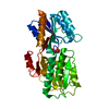
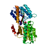
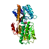
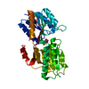
- PDB-1abf: SUBSTRATE SPECIFICITY AND AFFINITY OF A PROTEIN MODULATED BY BOUN... -
+ Open data

- Basic information
| Entry | Database: PDB / ID: 1abf | ||||||
|---|---|---|---|---|---|---|---|
| Title | SUBSTRATE SPECIFICITY AND AFFINITY OF A PROTEIN MODULATED BY BOUND WATER MOLECULES | ||||||
 Components Components | L-ARABINOSE-BINDING PROTEIN | ||||||
 Keywords Keywords | BINDING PROTEIN | ||||||
| Function / homology |  Function and homology information Function and homology informationABC-type monosaccharide transporter activity / L-arabinose transmembrane transport / monosaccharide binding / ATP-binding cassette (ABC) transporter complex, substrate-binding subunit-containing / outer membrane-bounded periplasmic space / carbohydrate binding / membrane Similarity search - Function | ||||||
| Biological species |  | ||||||
| Method |  X-RAY DIFFRACTION / Resolution: 1.9 Å X-RAY DIFFRACTION / Resolution: 1.9 Å | ||||||
 Authors Authors | Wilson, D.K. / Quiocho, F.A. | ||||||
 Citation Citation | Journal: Nature / Year: 1989 Title: Substrate specificity and affinity of a protein modulated by bound water molecules. Authors: Quiocho, F.A. / Wilson, D.K. / Vyas, N.K. #1: Journal: Nature / Year: 1984Title: Novel Stereospecificity of the L-Arabinose-Binding Protein Authors: Quiocho, F.A. / Vyas, N.K. | ||||||
| History |
|
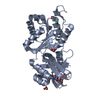
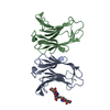
- Structure visualization
| Structure viewer | Molecule: MolmilJmol/JSmol |
|---|
- Downloads & links
-Download
| PDBx/mmCIF format | 1abf.cif.gz | 75.8 KB | Display | PDBx/mmCIF format |
|---|---|---|---|---|
| PDB format | pdb1abf.ent.gz | 55.5 KB | Display | PDB format |
| PDBx/mmJSON format | 1abf.json.gz | Tree view | PDBx/mmJSON format | |
| Others |  Other downloads Other downloads |
-Validation report
| Arichive directory | https://data.pdbj.org/pub/pdb/validation_reports/ab/1abfftp://data.pdbj.org/pub/pdb/validation_reports/ab/1abf | HTTPS FTP |
|---|
-Related structure data


-Links
 PDBj
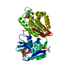
PDBj- Assembly
| Deposited unit | 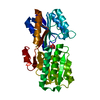
| ||||||||
|---|---|---|---|---|---|---|---|---|---|
| 1 |
| ||||||||
| Unit cell |
|
-Components
| #1: Protein | Mass: 33250.996 Da / Num. of mol.: 1 Source method: isolated from a genetically manipulated source Source: (gene. exp.) |
|---|---|
| #2: Sugar | ChemComp-FCA /   Type: D-saccharide, alpha linking / Mass: 164.156 Da / Num. of mol.: 1 Type: D-saccharide, alpha linking / Mass: 164.156 Da / Num. of mol.: 1Source method: isolated from a genetically manipulated source Formula: C6H12O5 |
| #3: Sugar | ChemComp-FCB /   Type: D-saccharide, beta linking / Mass: 164.156 Da / Num. of mol.: 1 Type: D-saccharide, beta linking / Mass: 164.156 Da / Num. of mol.: 1Source method: isolated from a genetically manipulated source Formula: C6H12O5 |
| #4: Water | ChemComp-HOH /  Mass: 18.015 Da / Num. of mol.: 191 / Source method: isolated from a natural source / Formula: H2O Mass: 18.015 Da / Num. of mol.: 191 / Source method: isolated from a natural source / Formula: H2O |
-Experimental details
-Experiment
| Experiment | Method: X-RAY DIFFRACTION |
|---|
- Sample preparation
| Crystal | Density Matthews: 2.36 Å3/Da / Density % sol: 47.91 % | |||||||||||||||
|---|---|---|---|---|---|---|---|---|---|---|---|---|---|---|---|---|
| Crystal grow | *PLUS pH: 6.5 / Method: microdialysis / Details: referred to J.Mol.Biol. 86.491-493 1974 | |||||||||||||||
| Components of the solutions | *PLUS
|
-Data collection
| Radiation | Scattering type: x-ray |
|---|---|
| Radiation wavelength | Relative weight: 1 |
- Processing
| Software | Name: PROLSQ / Classification: refinement | |||||||||||||||||||||||||||||||||||||||||||||||||||||||||||||||
|---|---|---|---|---|---|---|---|---|---|---|---|---|---|---|---|---|---|---|---|---|---|---|---|---|---|---|---|---|---|---|---|---|---|---|---|---|---|---|---|---|---|---|---|---|---|---|---|---|---|---|---|---|---|---|---|---|---|---|---|---|---|---|---|---|
| Refinement | Resolution: 1.9→10 Å /
| |||||||||||||||||||||||||||||||||||||||||||||||||||||||||||||||
| Refinement step | Cycle: LAST / Resolution: 1.9→10 Å
| |||||||||||||||||||||||||||||||||||||||||||||||||||||||||||||||
| Refine LS restraints |
| |||||||||||||||||||||||||||||||||||||||||||||||||||||||||||||||
| Refinement | *PLUS Highest resolution: 1.9 Å / Lowest resolution: 10 Å / Num. reflection obs: 18780 / Rfactor obs: 0.134 | |||||||||||||||||||||||||||||||||||||||||||||||||||||||||||||||
| Solvent computation | *PLUS | |||||||||||||||||||||||||||||||||||||||||||||||||||||||||||||||
| Displacement parameters | *PLUS | |||||||||||||||||||||||||||||||||||||||||||||||||||||||||||||||
| Refine LS restraints | *PLUS
|
