 Movie
Movie Controller


+ Open data

- Basic information
| Entry | Database: PDB / ID: 1a37 | ||||||
|---|---|---|---|---|---|---|---|
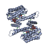
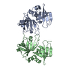
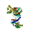
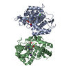
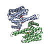
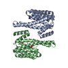
| Title | 14-3-3 PROTEIN ZETA BOUND TO PS-RAF259 PEPTIDE | ||||||
 Components Components |
| ||||||
 Keywords Keywords | COMPLEX (SIGNAL TRANSDUCTION/PEPTIDE) / SIGNAL TRANSDUCTION / COMPLEX (SIGNAL TRANSDUCTION-PEPTIDE) / COMPLEX (SIGNAL TRANSDUCTION-PEPTIDE) complex | ||||||
| Function / homology |  Function and homology information Function and homology informationKSRP (KHSRP) binds and destabilizes mRNA / RHO GTPases activate PKNs / Chk1/Chk2(Cds1) mediated inactivation of Cyclin B:Cdk1 complex / NOTCH4 Activation and Transmission of Signal to the Nucleus / Regulation of localization of FOXO transcription factors / Activation of BAD and translocation to mitochondria / GP1b-IX-V activation signalling / Deactivation of the beta-catenin transactivating complex / regulation of programmed cell death / synaptic target recognition ...KSRP (KHSRP) binds and destabilizes mRNA / RHO GTPases activate PKNs / Chk1/Chk2(Cds1) mediated inactivation of Cyclin B:Cdk1 complex / NOTCH4 Activation and Transmission of Signal to the Nucleus / Regulation of localization of FOXO transcription factors / Activation of BAD and translocation to mitochondria / GP1b-IX-V activation signalling / Deactivation of the beta-catenin transactivating complex / regulation of programmed cell death / synaptic target recognition / Rap1 signalling / TP53 Regulates Metabolic Genes / antibacterial innate immune response / Golgi reassembly / Interleukin-3, Interleukin-5 and GM-CSF signaling / establishment of Golgi localization / regulation of lysosome organization / respiratory system process / tube formation / regulation of synapse maturation / negative regulation of protein localization to nucleus / lysosome organization / TORC1 signaling / phosphoserine residue binding / regulation of ERK1 and ERK2 cascade / protein targeting / cellular response to glucose starvation / ERK1 and ERK2 cascade / negative regulation of TORC1 signaling / protein sequestering activity / lung development / negative regulation of innate immune response / hippocampal mossy fiber to CA3 synapse / intracellular protein localization / melanosome / angiogenesis / protein phosphatase binding / DNA-binding transcription factor binding / transmembrane transporter binding / protein phosphorylation / protein domain specific binding / ubiquitin protein ligase binding / protein kinase binding / glutamatergic synapse / negative regulation of transcription by RNA polymerase II / signal transduction / identical protein binding / nucleus / cytoplasm / cytosol Similarity search - Function | ||||||
| Biological species |  | ||||||
| Method |  X-RAY DIFFRACTION / MOLECULAR REPLACEMENT / Resolution: 3.6 Å X-RAY DIFFRACTION / MOLECULAR REPLACEMENT / Resolution: 3.6 Å | ||||||
 Authors Authors | Petosa, C. / Masters, S.C. / Pohl, J. / Wang, B. / Fu, H. / Liddington, R.C. | ||||||
 Citation Citation | Journal: J.Biol.Chem. / Year: 1998 Title: 14-3-3zeta binds a phosphorylated Raf peptide and an unphosphorylated peptide via its conserved amphipathic groove. Authors: Petosa, C. / Masters, S.C. / Bankston, L.A. / Pohl, J. / Wang, B. / Fu, H. / Liddington, R.C. #1: Journal: J.Biol.Chem. / Year: 1998Title: 14-3-3Zeta Binds a Phosphorylated Raf Peptide and an Unphosphorylated Peptide Via its Conserved Amphipathic Groove Authors: Petosa, C. / Masters, S.C. / Bankston, L.A. / Pohl, J. / Wang, B. / Fu, H. / Liddington, R.C. | ||||||
| History |
|
- Structure visualization
| Structure viewer | Molecule: MolmilJmol/JSmol |
|---|
- Downloads & links
-Download
| PDBx/mmCIF format | 1a37.cif.gz | 83.7 KB | Display | PDBx/mmCIF format |
|---|---|---|---|---|
| PDB format | pdb1a37.ent.gz | 65.2 KB | Display | PDB format |
| PDBx/mmJSON format | 1a37.json.gz | Tree view | PDBx/mmJSON format | |
| Others |  Other downloads Other downloads |
-Validation report
| Arichive directory | https://data.pdbj.org/pub/pdb/validation_reports/a3/1a37ftp://data.pdbj.org/pub/pdb/validation_reports/a3/1a37 | HTTPS FTP |
|---|
-Related structure data
-Links
 PDBj
PDBj


- Assembly
| Deposited unit | 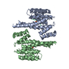
| ||||||||||||
|---|---|---|---|---|---|---|---|---|---|---|---|---|---|
| 1 |
| ||||||||||||
| Unit cell |
| ||||||||||||
| Noncrystallographic symmetry (NCS) | NCS oper:
|
-Components
| #1: Protein | Mass: 27777.092 Da / Num. of mol.: 2 Source method: isolated from a genetically manipulated source Details: COMPLEXED WITH PHOSPHOSERINE-CONTAINING PEPTIDE DERIVED FROM RAF Source: (gene. exp.)  #2: Protein/peptide | Mass: 1840.951 Da / Num. of mol.: 2 Source method: isolated from a genetically manipulated source Has protein modification | Y | |
|---|
-Experimental details
-Experiment
| Experiment | Method: X-RAY DIFFRACTION / Number of used crystals: 1 |
|---|
- Sample preparation
| Crystal | Density Matthews: 5.49 Å3/Da / Density % sol: 77.58 % | ||||||||||||||||||||||||||||||||||||
|---|---|---|---|---|---|---|---|---|---|---|---|---|---|---|---|---|---|---|---|---|---|---|---|---|---|---|---|---|---|---|---|---|---|---|---|---|---|
| Crystal grow | pH: 8.5 / Details: pH 8.5 | ||||||||||||||||||||||||||||||||||||
| Crystal grow | *PLUS Temperature: 4 ℃ / Method: vapor diffusion, hanging drop / Details: Liu, D., (1995) Nature, 376, 191. | ||||||||||||||||||||||||||||||||||||
| Components of the solutions | *PLUS
|
-Data collection
| Diffraction | Mean temperature: 100 K |
|---|---|
| Diffraction source | Source: ROTATING ANODE / Type: RIGAKU RUH2R / Wavelength: 1.5418 |
| Detector | Type: RIGAKU RAXIS IV / Detector: IMAGE PLATE / Date: Jun 1, 1997 / Details: MIRRORS |
| Radiation | Monochromator: NI FILTER / Monochromatic (M) / Laue (L): M / Scattering type: x-ray |
| Radiation wavelength | Wavelength: 1.5418 Å / Relative weight: 1 |
| Reflection | Resolution: 3.6→20 Å / Num. obs: 14700 / % possible obs: 99 % / Observed criterion σ(I): 0 / Redundancy: 4.3 % / Rsym value: 0.072 / Net I/σ(I): 20 |
| Reflection shell | Resolution: 3.6→3.8 Å / Redundancy: 3.7 % / Mean I/σ(I) obs: 7 / Rsym value: 0.258 / % possible all: 98.8 |
| Reflection | *PLUS Highest resolution: 3.6 Å / Lowest resolution: 20 Å / Num. obs: 14696 / % possible obs: 99.5 % / Rmerge(I) obs: 0.07 |
- Processing
| Software |
| ||||||||||||||||||||
|---|---|---|---|---|---|---|---|---|---|---|---|---|---|---|---|---|---|---|---|---|---|
| Refinement | Method to determine structure: MOLECULAR REPLACEMENT / Resolution: 3.6→20 Å / Data cutoff high absF: 10000000 / Data cutoff low absF: 0.001 / Cross valid method: THROUGHOUT / σ(F): 0
| ||||||||||||||||||||
| Refinement step | Cycle: LAST / Resolution: 3.6→20 Å
| ||||||||||||||||||||
| Software | *PLUS Name: X-PLOR / Version: 3.8 / Classification: refinement | ||||||||||||||||||||
| Refinement | *PLUS Lowest resolution: 20 Å / σ(F): 0 / Rfactor obs: 0.31 | ||||||||||||||||||||
| Solvent computation | *PLUS | ||||||||||||||||||||
| Displacement parameters | *PLUS | ||||||||||||||||||||
| Refine LS restraints | *PLUS
|
