 Movie
Movie Controller


+ Open data

- Basic information
| Entry | Database: PDB / ID: 1a0t | |||||||||
|---|---|---|---|---|---|---|---|---|---|---|
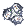
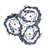
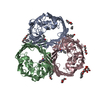
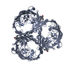
| Title | SUCROSE-SPECIFIC PORIN, WITH BOUND SUCROSE MOLECULES | |||||||||
 Components Components | SUCROSE-SPECIFIC PORIN | |||||||||
 Keywords Keywords | OUTER MEMBRANE PROTEIN / PORIN | |||||||||
| Function / homology |  Function and homology information Function and homology informationpolysaccharide transport / porin activity / pore complex / carbohydrate transmembrane transporter activity / monoatomic ion transport / cell outer membrane Similarity search - Function | |||||||||
| Biological species |  Salmonella typhimurium (bacteria) Salmonella typhimurium (bacteria) | |||||||||
| Method |  X-RAY DIFFRACTION / DIFFERENCE FOURIER / Resolution: 2.4 Å X-RAY DIFFRACTION / DIFFERENCE FOURIER / Resolution: 2.4 Å | |||||||||
 Authors Authors | Diederichs, K. / Welte, W. | |||||||||
 Citation Citation | Journal: Nat.Struct.Biol. / Year: 1998 Title: Structure of the sucrose-specific porin ScrY from Salmonella typhimurium and its complex with sucrose. Authors: Forst, D. / Welte, W. / Wacker, T. / Diederichs, K. #1: Journal: J.Mol.Biol. / Year: 1993Title: Crystallization and Preliminary X-Ray Diffraction Analysis of Scry, a Specific Bacterial Outer Membrane Porin Authors: Forst, D. / Schulein, K. / Wacker, T. / Diederichs, K. / Kreutz, W. / Benz, R. / Welte, W. | |||||||||
| History |
|
- Structure visualization
| Structure viewer | Molecule: MolmilJmol/JSmol |
|---|
- Downloads & links
-Download
| PDBx/mmCIF format | 1a0t.cif.gz | 254.4 KB | Display | PDBx/mmCIF format |
|---|---|---|---|---|
| PDB format | pdb1a0t.ent.gz | 204.1 KB | Display | PDB format |
| PDBx/mmJSON format | 1a0t.json.gz | Tree view | PDBx/mmJSON format | |
| Others |  Other downloads Other downloads |
-Validation report
| Arichive directory | https://data.pdbj.org/pub/pdb/validation_reports/a0/1a0tftp://data.pdbj.org/pub/pdb/validation_reports/a0/1a0t | HTTPS FTP |
|---|
-Related structure data
| Related structure data |  1a0sC  1aosS S: Starting model for refinement C: citing same article ( |
|---|---|
| Similar structure data |


-Links
 PDBj
PDBj- Assembly
| Deposited unit | 
| |||||||||||||||||||||
|---|---|---|---|---|---|---|---|---|---|---|---|---|---|---|---|---|---|---|---|---|---|---|
| 1 |
| |||||||||||||||||||||
| Unit cell |
| |||||||||||||||||||||
| Noncrystallographic symmetry (NCS) | NCS domain:
NCS oper:
|
-Components
| #1: Protein | Mass: 45333.668 Da / Num. of mol.: 3 Source method: isolated from a genetically manipulated source Source: (gene. exp.) Salmonella typhimurium (bacteria) / Cellular location: OUTER MEMBRANE / Gene: SCRY / Plasmid: PSO112 / Cellular location (production host): OUTER MEMBRANE / Production host: #2: Polysaccharide | beta-D-fructofuranose-(2-1)-alpha-D-glucopyranose / sucrose   Source method: isolated from a genetically manipulated source Details: oligosaccharide with reducing-end-to-reducing-end glycosidic bond References: sucrose #3: Chemical |   Mass: 40.078 Da / Num. of mol.: 3 / Source method: obtained synthetically / Formula: Ca Mass: 40.078 Da / Num. of mol.: 3 / Source method: obtained synthetically / Formula: Ca#4: Water | ChemComp-HOH / |  Mass: 18.015 Da / Num. of mol.: 330 / Source method: isolated from a natural source / Formula: H2O Mass: 18.015 Da / Num. of mol.: 330 / Source method: isolated from a natural source / Formula: H2O |
|---|
-Experimental details
-Experiment
| Experiment | Method: X-RAY DIFFRACTION / Number of used crystals: 1 |
|---|
- Sample preparation
| Crystal | Density Matthews: 3.8 Å3/Da / Density % sol: 68 % | ||||||||||||||||||||||||||||||||||||||||||||||||||||||||||||||||||
|---|---|---|---|---|---|---|---|---|---|---|---|---|---|---|---|---|---|---|---|---|---|---|---|---|---|---|---|---|---|---|---|---|---|---|---|---|---|---|---|---|---|---|---|---|---|---|---|---|---|---|---|---|---|---|---|---|---|---|---|---|---|---|---|---|---|---|---|
| Crystal grow | Method: vapor diffusion, sitting drop / pH: 7.7 Details: PROTEIN WAS CRYSTALLIZED BY VAPOR DIFFUSION USING THE SITTING-DROP METHOD. THE DROP CONTAINED 5-7 MG/ML PROTEIN, 20 MM TRIS/CL AT PH 7.7, 100MM LICL, 20MM MGSO4, 1.2% BETA-D- ...Details: PROTEIN WAS CRYSTALLIZED BY VAPOR DIFFUSION USING THE SITTING-DROP METHOD. THE DROP CONTAINED 5-7 MG/ML PROTEIN, 20 MM TRIS/CL AT PH 7.7, 100MM LICL, 20MM MGSO4, 1.2% BETA-D-OCTYLGLUCOPYRANOSIDE AND 6-9% PEG-2000. THE CONCENTRATION OF PEG IN THE RESERVOIR WAS 12-15%. 2M SUCROSE WAS ADDED TO THE DROP FOR COCRYSTALLIZATION., vapor diffusion - sitting drop | ||||||||||||||||||||||||||||||||||||||||||||||||||||||||||||||||||
| Crystal grow | *PLUS Temperature: 17 ℃ / Method: vapor diffusion, sitting drop | ||||||||||||||||||||||||||||||||||||||||||||||||||||||||||||||||||
| Components of the solutions | *PLUS
|
-Data collection
| Diffraction | Mean temperature: 287 K |
|---|---|
| Diffraction source | Type: OTHER / Wavelength: 1.5418 |
| Detector | Type: STOE / Detector: DIFFRACTOMETER / Date: Apr 1, 1995 / Details: COLLIMATOR |
| Radiation | Monochromator: GRAPHITE(002) / Monochromatic (M) / Laue (L): M / Scattering type: x-ray |
| Radiation wavelength | Wavelength: 1.5418 Å / Relative weight: 1 |
| Reflection | Resolution: 2.4→17 Å / Num. obs: 65815 / % possible obs: 80.7 % / Observed criterion σ(I): 0 / Redundancy: 2.74 % / Biso Wilson estimate: 19.4 Å2 / Rsym value: 0.155 / Net I/σ(I): 5 |
| Reflection shell | Resolution: 2.4→3 Å / Redundancy: 2.6 % / Mean I/σ(I) obs: 2 / Rsym value: 0.524 / % possible all: 84 |
| Reflection | *PLUS Rmerge(I) obs: 0.161 |
- Processing
| Software |
| ||||||||||||||||||||||||||||||||||||||||||||||||||||||||||||||||||||||||||||||||
|---|---|---|---|---|---|---|---|---|---|---|---|---|---|---|---|---|---|---|---|---|---|---|---|---|---|---|---|---|---|---|---|---|---|---|---|---|---|---|---|---|---|---|---|---|---|---|---|---|---|---|---|---|---|---|---|---|---|---|---|---|---|---|---|---|---|---|---|---|---|---|---|---|---|---|---|---|---|---|---|---|---|
| Refinement | Method to determine structure: DIFFERENCE FOURIER Starting model: SUCROSE PORIN (PDB ENTRY 1AOS) Resolution: 2.4→100 Å / Rfactor Rfree error: 0.008 / Data cutoff high absF: 100000 / Data cutoff low absF: 0.1 / Isotropic thermal model: RESTRAINED / Cross valid method: THROUGHOUT / σ(F): 1
| ||||||||||||||||||||||||||||||||||||||||||||||||||||||||||||||||||||||||||||||||
| Displacement parameters | Biso mean: 28.4 Å2 | ||||||||||||||||||||||||||||||||||||||||||||||||||||||||||||||||||||||||||||||||
| Refine analyze |
| ||||||||||||||||||||||||||||||||||||||||||||||||||||||||||||||||||||||||||||||||
| Refinement step | Cycle: LAST / Resolution: 2.4→100 Å
| ||||||||||||||||||||||||||||||||||||||||||||||||||||||||||||||||||||||||||||||||
| Refine LS restraints |
| ||||||||||||||||||||||||||||||||||||||||||||||||||||||||||||||||||||||||||||||||
| Refine LS restraints NCS | Refine-ID: X-RAY DIFFRACTION / Rms dev position: 0.1 Å / Weight Biso : 1 / Weight position: 50
| ||||||||||||||||||||||||||||||||||||||||||||||||||||||||||||||||||||||||||||||||
| LS refinement shell | Resolution: 2.4→2.49 Å / Rfactor Rfree error: 0.058 / Total num. of bins used: 10
| ||||||||||||||||||||||||||||||||||||||||||||||||||||||||||||||||||||||||||||||||
| Xplor file |
| ||||||||||||||||||||||||||||||||||||||||||||||||||||||||||||||||||||||||||||||||
| Software | *PLUS Name: X-PLOR / Version: 3.851 / Classification: refinement | ||||||||||||||||||||||||||||||||||||||||||||||||||||||||||||||||||||||||||||||||
| Refine LS restraints | *PLUS
|
