 Movie
Movie Controller

[English] 日本語
 Yorodumi
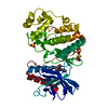
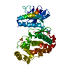
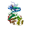
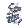
- PDB-6fle: Crystal structure of ERK2 in complex with an adenosine derivative -
+ Open data

- Basic information
| Entry | Database: PDB / ID: 6fle | ||||||
|---|---|---|---|---|---|---|---|
| Title | Crystal structure of ERK2 in complex with an adenosine derivative | ||||||
 Components Components | Mitogen-activated protein kinase 1 | ||||||
 Keywords Keywords |  TRANSFERASE / SERINE/THREONINE-PROTEIN KINASE TRANSFERASE / SERINE/THREONINE-PROTEIN KINASE | ||||||
| Function / homology |  Function and homology information Function and homology informationphospho-PLA2 pathway / RAF-independent MAPK1/3 activation / MAPK1 (ERK2) activation / Signaling by NODAL / Frs2-mediated activation / ERK/MAPK targets / ERKs are inactivated / Activation of the AP-1 family of transcription factors / RHO GTPases Activate WASPs and WAVEs / IFNG signaling activates MAPKs ...phospho-PLA2 pathway / RAF-independent MAPK1/3 activation / MAPK1 (ERK2) activation / Signaling by NODAL / Frs2-mediated activation / ERK/MAPK targets / ERKs are inactivated / Activation of the AP-1 family of transcription factors / RHO GTPases Activate WASPs and WAVEs / IFNG signaling activates MAPKs / Negative feedback regulation of MAPK pathway / Gastrin-CREB signalling pathway via PKC and MAPK / Estrogen-dependent nuclear events downstream of ESR-membrane signaling / Golgi Cisternae Pericentriolar Stack Reorganization / Regulation of actin dynamics for phagocytic cup formation / Estrogen-stimulated signaling through PRKCZ / Growth hormone receptor signaling / Spry regulation of FGF signaling / SMAD2/SMAD3:SMAD4 heterotrimer regulates transcription / Oxidative Stress Induced Senescence / Senescence-Associated Secretory Phenotype (SASP) / Oncogene Induced Senescence / Signaling by Activin / Downregulation of SMAD2/3:SMAD4 transcriptional activity / Signal attenuation / NCAM signaling for neurite out-growth / Negative regulation of FGFR1 signaling / Negative regulation of FGFR3 signaling / Negative regulation of FGFR4 signaling / ESR-mediated signaling / Regulation of the apoptosome activity / Interferon gamma signaling / Signal transduction by L1 / Negative regulation of FGFR2 signaling / RHO GTPases Activate NADPH Oxidases / Negative regulation of MAPK pathway / FCERI mediated MAPK activation / PI5P, PP2A and IER3 Regulate PI3K/AKT Signaling / Regulation of HSF1-mediated heat shock response / MAP2K and MAPK activation / diadenosine tetraphosphate biosynthetic process / Recycling pathway of L1 / neural crest cell development / cardiac neural crest cell development involved in heart development / caveolin-mediated endocytosis / cytosine metabolic process / response to epidermal growth factor / mitogen-activated protein kinase kinase kinase binding / regulation of cellular pH / positive regulation of macrophage proliferation / outer ear morphogenesis / Thrombin signalling through proteinase activated receptors (PARs) / RAF/MAP kinase cascade / regulation of Golgi inheritance / ERBB signaling pathway / labyrinthine layer blood vessel development / mammary gland epithelial cell proliferation / Neutrophil degranulation / trachea formation / regulation of cytoskeleton organization / regulation of early endosome to late endosome transport / cellular response to organic substance / regulation of stress-activated MAPK cascade / positive regulation of macrophage chemotaxis / response to exogenous dsRNA / lung morphogenesis / ERBB2-ERBB3 signaling pathway / face development / androgen receptor signaling pathway / pseudopodium / progesterone receptor signaling pathway / positive regulation of telomere capping / negative regulation of cell differentiation / Bergmann glial cell differentiation / thyroid gland development / decidualization / steroid hormone mediated signaling pathway / regulation of ossification / MAP kinase activity / phosphatase binding / mitogen-activated protein kinase / stress-activated MAPK cascade / Schwann cell development / sensory perception of pain / lipopolysaccharide-mediated signaling pathway / positive regulation of cardiac muscle cell proliferation / ERK1 and ERK2 cascade / cellular response to cadmium ion / positive regulation of telomere maintenance via telomerase / cellular response to amino acid starvation / myelination / dendrite cytoplasm / phosphotyrosine residue binding / RNA polymerase II CTD heptapeptide repeat kinase activity / insulin-like growth factor receptor signaling pathway / positive regulation of translation / thymus development / positive regulation of peptidyl-threonine phosphorylation / response to nicotine / long-term synaptic potentiationSimilarity search - Function | ||||||
| Biological species |  Rattus norvegicus (Norway rat) Rattus norvegicus (Norway rat) | ||||||
| Method | X-RAY DIFFRACTION / SYNCHROTRON / MOLECULAR REPLACEMENT / Resolution: 1.48 Å | ||||||
 Authors Authors | Gelin, M. / Labesse, G. | ||||||
| Funding support |  France, 1items France, 1items
| ||||||
 Citation Citation | Journal: To be published Title: Crystal structure of ERK2 in complex with an adenosine derivative Authors: Gelin, M. / Pochet, S. / Labesse, G. | ||||||
| History |
|
- Structure visualization
| Structure viewer | Molecule: MolmilJmol/JSmol |
|---|
- Downloads & links
-Download
| PDBx/mmCIF format | 6fle.cif.gz | 171.6 KB | Display | PDBx/mmCIF format |
|---|---|---|---|---|
| PDB format | pdb6fle.ent.gz | 133.9 KB | Display | PDB format |
| PDBx/mmJSON format | 6fle.json.gz | Tree view | PDBx/mmJSON format | |
| Others |  Other downloads Other downloads |
-Validation report
| Arichive directory | https://data.pdbj.org/pub/pdb/validation_reports/fl/6fleftp://data.pdbj.org/pub/pdb/validation_reports/fl/6fle | HTTPS FTP |
|---|
-Related structure data
| Related structure data |  6fi3C  6fi6C  6fj0C  6fjbC  6fjzC  3qyzS S: Starting model for refinement C: citing same article ( |
|---|---|
| Similar structure data |


-Links
 PDBj
PDBj


- Assembly
| Deposited unit | 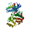
| ||||||||
|---|---|---|---|---|---|---|---|---|---|
| 1 |
| ||||||||
| Unit cell |
|
-Components
| #1: Protein | Mass: 42235.547 Da / Num. of mol.: 1 Source method: isolated from a genetically manipulated source Source: (gene. exp.) Rattus norvegicus (Norway rat) / Gene: Mapk1, Erk2, Mapk, Prkm1 / Production host:  Escherichia coli (E. coli) Escherichia coli (E. coli)References: UniProt: P63086, mitogen-activated protein kinase |
|---|---|
| #2: Chemical | ChemComp-SO4 / Sulfate  Mass: 96.063 Da / Num. of mol.: 1 / Source method: obtained synthetically / Formula: SO4 Mass: 96.063 Da / Num. of mol.: 1 / Source method: obtained synthetically / Formula: SO4 |
| #3: Chemical | ChemComp-DQ2 / [(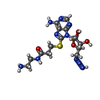  Mass: 439.473 Da / Num. of mol.: 1 / Source method: obtained synthetically / Formula: C15H23N10O4S Mass: 439.473 Da / Num. of mol.: 1 / Source method: obtained synthetically / Formula: C15H23N10O4S |
| #4: Chemical | ChemComp-DMS / Dimethyl sulfoxide  Mass: 78.133 Da / Num. of mol.: 1 / Source method: obtained synthetically / Formula: C2H6OS / Comment: DMSO, precipitant*YM Mass: 78.133 Da / Num. of mol.: 1 / Source method: obtained synthetically / Formula: C2H6OS / Comment: DMSO, precipitant*YM |
| #5: Water | ChemComp-HOH / Water Mass: 18.015 Da / Num. of mol.: 293 / Source method: isolated from a natural source / Formula: H2O Mass: 18.015 Da / Num. of mol.: 293 / Source method: isolated from a natural source / Formula: H2O |
-Experimental details
-Experiment
| Experiment | Method: X-RAY DIFFRACTION / Number of used crystals: 1 |
|---|
- Sample preparation
| Crystal | Density Matthews: 2.29 Å3/Da / Density % sol: 46.2 % |
|---|---|
| Crystal grow | Temperature: 291 K / Method: vapor diffusion, hanging drop / pH: 6.5 Details: PEG MME 2000, 0.1M MES pH 6.5, 0.1M ammonium sulfate, 0.02M beta-mercaptoethanol, 0.002M magnesium sulfate |
-Data collection
| Diffraction | Mean temperature: 100 K | ||||||||||||||||||||||||||||||||||||||||||||||||||||||||||||||||||||||||||||||||||||||||||||||||||||||||||||||||||||||||||||||||||||||||||||||||||||||||||||||||||||||||||||||||||||||||||||||||||||||||||||||||||
|---|---|---|---|---|---|---|---|---|---|---|---|---|---|---|---|---|---|---|---|---|---|---|---|---|---|---|---|---|---|---|---|---|---|---|---|---|---|---|---|---|---|---|---|---|---|---|---|---|---|---|---|---|---|---|---|---|---|---|---|---|---|---|---|---|---|---|---|---|---|---|---|---|---|---|---|---|---|---|---|---|---|---|---|---|---|---|---|---|---|---|---|---|---|---|---|---|---|---|---|---|---|---|---|---|---|---|---|---|---|---|---|---|---|---|---|---|---|---|---|---|---|---|---|---|---|---|---|---|---|---|---|---|---|---|---|---|---|---|---|---|---|---|---|---|---|---|---|---|---|---|---|---|---|---|---|---|---|---|---|---|---|---|---|---|---|---|---|---|---|---|---|---|---|---|---|---|---|---|---|---|---|---|---|---|---|---|---|---|---|---|---|---|---|---|---|---|---|---|---|---|---|---|---|---|---|---|---|---|---|---|---|
| Diffraction source | Source: SYNCHROTRON / Site: ESRF / Beamline: MASSIF-3 / Wavelength: 0.9677 Å | ||||||||||||||||||||||||||||||||||||||||||||||||||||||||||||||||||||||||||||||||||||||||||||||||||||||||||||||||||||||||||||||||||||||||||||||||||||||||||||||||||||||||||||||||||||||||||||||||||||||||||||||||||
| Detector | Type: DECTRIS EIGER X 4M / Detector: PIXEL / Date: Jul 25, 2016 | ||||||||||||||||||||||||||||||||||||||||||||||||||||||||||||||||||||||||||||||||||||||||||||||||||||||||||||||||||||||||||||||||||||||||||||||||||||||||||||||||||||||||||||||||||||||||||||||||||||||||||||||||||
| Radiation | Protocol: SINGLE WAVELENGTH / Monochromatic (M) / Laue (L): M / Scattering type: x-ray | ||||||||||||||||||||||||||||||||||||||||||||||||||||||||||||||||||||||||||||||||||||||||||||||||||||||||||||||||||||||||||||||||||||||||||||||||||||||||||||||||||||||||||||||||||||||||||||||||||||||||||||||||||
| Radiation wavelength | Wavelength: 0.9677 Å / Relative weight: 1 | ||||||||||||||||||||||||||||||||||||||||||||||||||||||||||||||||||||||||||||||||||||||||||||||||||||||||||||||||||||||||||||||||||||||||||||||||||||||||||||||||||||||||||||||||||||||||||||||||||||||||||||||||||
| Reflection | Resolution: 1.48→38.571 Å / Num. obs: 59645 / % possible obs: 94.1 % / Redundancy: 5.609 % / Biso Wilson estimate: 23.05 Å2 / CC1/2: 0.999 / Rmerge(I) obs: 0.038 / Rrim(I) all: 0.042 / Χ2: 0.994 / Net I/σ(I): 18.99 / Num. measured all: 334576 | ||||||||||||||||||||||||||||||||||||||||||||||||||||||||||||||||||||||||||||||||||||||||||||||||||||||||||||||||||||||||||||||||||||||||||||||||||||||||||||||||||||||||||||||||||||||||||||||||||||||||||||||||||
| Reflection shell | Diffraction-ID: 1
|
- Processing
| Software |
| ||||||||||||||||||||||||||||||||||||||||||||||||||||||||||||||||||||||||||||||||||||||||||||||||||||||||||||||||||||||||||||||||||||||||||||||||||||||||||
|---|---|---|---|---|---|---|---|---|---|---|---|---|---|---|---|---|---|---|---|---|---|---|---|---|---|---|---|---|---|---|---|---|---|---|---|---|---|---|---|---|---|---|---|---|---|---|---|---|---|---|---|---|---|---|---|---|---|---|---|---|---|---|---|---|---|---|---|---|---|---|---|---|---|---|---|---|---|---|---|---|---|---|---|---|---|---|---|---|---|---|---|---|---|---|---|---|---|---|---|---|---|---|---|---|---|---|---|---|---|---|---|---|---|---|---|---|---|---|---|---|---|---|---|---|---|---|---|---|---|---|---|---|---|---|---|---|---|---|---|---|---|---|---|---|---|---|---|---|---|---|---|---|---|---|---|
| Refinement | Method to determine structure: MOLECULAR REPLACEMENT Starting model: 3QYZ Resolution: 1.48→38.571 Å / SU ML: 0.15 / Cross valid method: THROUGHOUT / σ(F): 1.36 / Phase error: 19.01 / Stereochemistry target values: ML
| ||||||||||||||||||||||||||||||||||||||||||||||||||||||||||||||||||||||||||||||||||||||||||||||||||||||||||||||||||||||||||||||||||||||||||||||||||||||||||
| Solvent computation | Shrinkage radii: 0.9 Å / VDW probe radii: 1.11 Å / Solvent model: FLAT BULK SOLVENT MODEL | ||||||||||||||||||||||||||||||||||||||||||||||||||||||||||||||||||||||||||||||||||||||||||||||||||||||||||||||||||||||||||||||||||||||||||||||||||||||||||
| Displacement parameters | Biso max: 107.38 Å2 / Biso mean: 36.0822 Å2 / Biso min: 14.85 Å2 | ||||||||||||||||||||||||||||||||||||||||||||||||||||||||||||||||||||||||||||||||||||||||||||||||||||||||||||||||||||||||||||||||||||||||||||||||||||||||||
| Refinement step | Cycle: final / Resolution: 1.48→38.571 Å
| ||||||||||||||||||||||||||||||||||||||||||||||||||||||||||||||||||||||||||||||||||||||||||||||||||||||||||||||||||||||||||||||||||||||||||||||||||||||||||
| LS refinement shell | Refine-ID: X-RAY DIFFRACTION / Rfactor Rfree error: 0 / Total num. of bins used: 21
|
