 Movie
Movie Controller

[English] 日本語
 Yorodumi
- PDB-3tc5: Selective targeting of disease-relevant protein binding domains b... -
+ Open data

- Basic information
| Entry | Database: PDB / ID: 3tc5 | ||||||
|---|---|---|---|---|---|---|---|
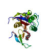
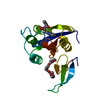
| Title | Selective targeting of disease-relevant protein binding domains by O-phosphorylated natural product derivatives | ||||||
 Components Components | Peptidyl-prolyl cis-trans isomerase NIMA-interacting 1 | ||||||
 Keywords Keywords | ISOMERASE/ISOMERASE INHIBITOR / PIN1 mutant (R14A) / Oncogenic transformation /  Small molecule / Cell cycle / Rotamase / Phosphoprotein / Nucleus / ISOMERASE-ISOMERASE INHIBITOR complex Small molecule / Cell cycle / Rotamase / Phosphoprotein / Nucleus / ISOMERASE-ISOMERASE INHIBITOR complex | ||||||
| Function / homology |  Function and homology informationcis-trans isomerase activity / phosphothreonine residue binding / negative regulation of cell motility / ubiquitin ligase activator activity / regulation of protein localization to nucleus / GTPase activating protein binding / postsynaptic cytosol / mitogen-activated protein kinase kinase binding / regulation of mitotic nuclear division / negative regulation of amyloid-beta formation ...cis-trans isomerase activity / phosphothreonine residue binding / negative regulation of cell motility / ubiquitin ligase activator activity / regulation of protein localization to nucleus / GTPase activating protein binding / postsynaptic cytosol / mitogen-activated protein kinase kinase binding / regulation of mitotic nuclear division / negative regulation of amyloid-beta formation / negative regulation of SMAD protein signal transduction / PI5P Regulates TP53 Acetylation / cytoskeletal motor activity / RHO GTPases Activate NADPH Oxidases / phosphoserine residue binding / protein peptidyl-prolyl isomerization / positive regulation of protein dephosphorylation / ciliary basal body / negative regulation of protein binding / regulation of cytokinesis / peptidylprolyl isomerase / peptidyl-prolyl cis-trans isomerase activity / Negative regulators of DDX58/IFIH1 signaling / synapse organization / phosphoprotein binding / regulation of protein phosphorylation / negative regulation of transforming growth factor beta receptor signaling pathway / tau protein binding / regulation of protein stability / negative regulation of protein catabolic process / negative regulation of ERK1 and ERK2 cascade / ISG15 antiviral mechanism / neuron differentiation / beta-catenin binding / positive regulation of GTPase activity / positive regulation of canonical Wnt signaling pathway / positive regulation of protein binding / midbody / regulation of gene expression / Regulation of TP53 Activity through Phosphorylation / response to hypoxia / protein stabilization / nuclear speck / cell cycle / positive regulation of protein phosphorylation / glutamatergic synapse / positive regulation of transcription by RNA polymerase II / nucleoplasm / nucleus / cytosol / cytoplasm Function and homology informationcis-trans isomerase activity / phosphothreonine residue binding / negative regulation of cell motility / ubiquitin ligase activator activity / regulation of protein localization to nucleus / GTPase activating protein binding / postsynaptic cytosol / mitogen-activated protein kinase kinase binding / regulation of mitotic nuclear division / negative regulation of amyloid-beta formation ...cis-trans isomerase activity / phosphothreonine residue binding / negative regulation of cell motility / ubiquitin ligase activator activity / regulation of protein localization to nucleus / GTPase activating protein binding / postsynaptic cytosol / mitogen-activated protein kinase kinase binding / regulation of mitotic nuclear division / negative regulation of amyloid-beta formation / negative regulation of SMAD protein signal transduction / PI5P Regulates TP53 Acetylation / cytoskeletal motor activity / RHO GTPases Activate NADPH Oxidases / phosphoserine residue binding / protein peptidyl-prolyl isomerization / positive regulation of protein dephosphorylation / ciliary basal body / negative regulation of protein binding / regulation of cytokinesis / peptidylprolyl isomerase / peptidyl-prolyl cis-trans isomerase activity / Negative regulators of DDX58/IFIH1 signaling / synapse organization / phosphoprotein binding / regulation of protein phosphorylation / negative regulation of transforming growth factor beta receptor signaling pathway / tau protein binding / regulation of protein stability / negative regulation of protein catabolic process / negative regulation of ERK1 and ERK2 cascade / ISG15 antiviral mechanism / neuron differentiation / beta-catenin binding / positive regulation of GTPase activity / positive regulation of canonical Wnt signaling pathway / positive regulation of protein binding / midbody / regulation of gene expression / Regulation of TP53 Activity through Phosphorylation / response to hypoxia / protein stabilization / nuclear speck / cell cycle / positive regulation of protein phosphorylation / glutamatergic synapse / positive regulation of transcription by RNA polymerase II / nucleoplasm / nucleus / cytosol / cytoplasmSimilarity search - Function | ||||||
| Biological species |  Homo sapiens (human) Homo sapiens (human) | ||||||
| Method | X-RAY DIFFRACTION / SYNCHROTRON / MOLECULAR REPLACEMENT / Resolution: 1.4 Å | ||||||
 Authors Authors | Graeber, M. / Janczyk, W. / Sperl, B. / Elumalai, N. / Kozany, C. / Hausch, F. / Holak, T.A. / Berg, T. | ||||||
 Citation Citation | Journal: Acs Chem.Biol. / Year: 2011 Title: Selective targeting of disease-relevant protein binding domains by o-phosphorylated natural product derivatives. Authors: Graber, M. / Janczyk, W. / Sperl, B. / Elumalai, N. / Kozany, C. / Hausch, F. / Holak, T.A. / Berg, T. | ||||||
| History |
|
- Structure visualization
| Structure viewer | Molecule: MolmilJmol/JSmol |
|---|
- Downloads & links
-Download
| PDBx/mmCIF format | 3tc5.cif.gz | 84.6 KB | Display | PDBx/mmCIF format |
|---|---|---|---|---|
| PDB format | pdb3tc5.ent.gz | 62.5 KB | Display | PDB format |
| PDBx/mmJSON format | 3tc5.json.gz | Tree view | PDBx/mmJSON format | |
| Others |  Other downloads Other downloads |
-Validation report
| Arichive directory | https://data.pdbj.org/pub/pdb/validation_reports/tc/3tc5ftp://data.pdbj.org/pub/pdb/validation_reports/tc/3tc5 | HTTPS FTP |
|---|
-Related structure data
| Related structure data |  2zr6S S: Starting model for refinement |
|---|---|
| Similar structure data |


-Links
 PDBj
PDBj


- Assembly
| Deposited unit | 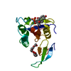
| ||||||||
|---|---|---|---|---|---|---|---|---|---|
| 1 |
| ||||||||
| Unit cell |
|
-Components
| #1: Protein | Mass: 18467.473 Da / Num. of mol.: 1 Fragment: Peptidyl-prolyl cis-trans isomerase NIMA-interacting 1 Mutation: R14A Source method: isolated from a genetically manipulated source Source: (gene. exp.) Homo sapiens (human) / Gene: PIN1 / Production host:  Escherichia coli (E. coli) / References: UniProt: Q13526, peptidylprolyl isomerase Escherichia coli (E. coli) / References: UniProt: Q13526, peptidylprolyl isomerase |
|---|---|
| #2: Chemical | ChemComp-3T5 / (  Mass: 472.441 Da / Num. of mol.: 1 / Source method: obtained synthetically / Formula: C22H30FO8P Mass: 472.441 Da / Num. of mol.: 1 / Source method: obtained synthetically / Formula: C22H30FO8P |
| #3: Chemical | ChemComp-P6G / Polyethylene glycol  Mass: 282.331 Da / Num. of mol.: 1 / Source method: obtained synthetically / Formula: C12H26O7 / Comment: precipitant*YM Mass: 282.331 Da / Num. of mol.: 1 / Source method: obtained synthetically / Formula: C12H26O7 / Comment: precipitant*YM |
| #4: Water | ChemComp-HOH / Water Mass: 18.015 Da / Num. of mol.: 266 / Source method: isolated from a natural source / Formula: H2O Mass: 18.015 Da / Num. of mol.: 266 / Source method: isolated from a natural source / Formula: H2O |
-Experimental details
-Experiment
| Experiment | Method: X-RAY DIFFRACTION / Number of used crystals: 1 |
|---|
- Sample preparation
| Crystal | Density Matthews: 2.9 Å3/Da / Density % sol: 57.57 % |
|---|---|
| Crystal grow | Temperature: 277 K / Method: vapor diffusion, sitting drop / pH: 7.5 Details: 2.2M Ammonium sulphate, 0.1M HEPES buffer, 1% PEG 400,pH 7.5, vapor diffusion, sitting drop, temperature 277K, VAPOR DIFFUSION, SITTING DROP |
-Data collection
| Diffraction | Mean temperature: 77 K |
|---|---|
| Diffraction source | Source: SYNCHROTRON / Site: SLS  / Beamline: X10SA / Wavelength: 1 Å / Beamline: X10SA / Wavelength: 1 Å |
| Detector | Type: PSI PILATUS 6M / Detector: PIXEL / Date: Oct 18, 2010 |
| Radiation | Monochromator: mirror / Protocol: SINGLE WAVELENGTH / Monochromatic (M) / Laue (L): M / Scattering type: x-ray |
| Radiation wavelength | Wavelength: 1 Å / Relative weight: 1 |
| Reflection | Resolution: 1.4→59 Å / Num. all: 42748 / Num. obs: 42028 / % possible obs: 98.3 % / Observed criterion σ(I): -3 |
| Reflection shell | Resolution: 1.4→1.7 Å / Rmerge(I) obs: 0.338 / Mean I/σ(I) obs: 4.7 / % possible all: 96.9 |
- Processing
| Software |
| ||||||||||||||||||||||||||||||||||||||||||||||||||||||||||||||||||||||||||||||||||||||||||
|---|---|---|---|---|---|---|---|---|---|---|---|---|---|---|---|---|---|---|---|---|---|---|---|---|---|---|---|---|---|---|---|---|---|---|---|---|---|---|---|---|---|---|---|---|---|---|---|---|---|---|---|---|---|---|---|---|---|---|---|---|---|---|---|---|---|---|---|---|---|---|---|---|---|---|---|---|---|---|---|---|---|---|---|---|---|---|---|---|---|---|---|
| Refinement | Method to determine structure: MOLECULAR REPLACEMENT Starting model: 2ZR6 Resolution: 1.4→19.73 Å / Cor.coef. Fo:Fc: 0.965 / Cor.coef. Fo:Fc free: 0.944 / SU B: 2.064 / SU ML: 0.037 / Cross valid method: THROUGHOUT / ESU R Free: 0.069 / Stereochemistry target values: MAXIMUM LIKELIHOOD / Details: HYDROGENS HAVE BEEN ADDED IN THE RIDING POSITIONS
| ||||||||||||||||||||||||||||||||||||||||||||||||||||||||||||||||||||||||||||||||||||||||||
| Solvent computation | Ion probe radii: 0.8 Å / Shrinkage radii: 0.8 Å / VDW probe radii: 1.4 Å / Solvent model: MASK | ||||||||||||||||||||||||||||||||||||||||||||||||||||||||||||||||||||||||||||||||||||||||||
| Displacement parameters | Biso mean: 15.45 Å2
| ||||||||||||||||||||||||||||||||||||||||||||||||||||||||||||||||||||||||||||||||||||||||||
| Refinement step | Cycle: LAST / Resolution: 1.4→19.73 Å
| ||||||||||||||||||||||||||||||||||||||||||||||||||||||||||||||||||||||||||||||||||||||||||
| Refine LS restraints |
| ||||||||||||||||||||||||||||||||||||||||||||||||||||||||||||||||||||||||||||||||||||||||||
| LS refinement shell | Resolution: 1.4→1.7 Å / Total num. of bins used: 20
|
