Resolution: 1.8→24 Å / Num. obs: 54988 / % possible obs: 92.5 % / Redundancy: 7.2 % / Rmerge(I) obs: 0.083 / Net I/σ(I): 13.2
Reflection shell
Resolution: 1.8→1.83 Å / Redundancy: 3.46 % / Rmerge(I) obs: 0.207 / Mean I/σ(I) obs: 6.29 / % possible all: 94
-
Processing
Software
Name
Classification
DENZO
datareduction
SCALEPACK
datascaling
SHELXL-97
phasing
SOLVE
phasing
SHELXL-97
refinement
Refinement
Method to determine structure: MAD / Resolution: 1.8→24 Å / Num. parameters: 20171 / Num. restraintsaints: 18190 / Cross valid method: FREE R-VALUE / σ(F): 0 / Stereochemistry target values: ENGH AND HUBER Details: SER 113 HAS ONLY PARTIAL DENSITY AND THIS IS RESPONSIBLE FOR THE POOR QUALITY OF GLU A 114. RESIDUE ARG A 487 SHOWS POOR DENSITY FOR ATOMS CG AND CD. DATA WAS COLLECTED AT WAVELENGTHS 1.736 ...Details: SER 113 HAS ONLY PARTIAL DENSITY AND THIS IS RESPONSIBLE FOR THE POOR QUALITY OF GLU A 114. RESIDUE ARG A 487 SHOWS POOR DENSITY FOR ATOMS CG AND CD. DATA WAS COLLECTED AT WAVELENGTHS 1.736 AND 1.739 ANGSTROM USING THE EMBL/DESY, HAMBURG BEAMLINE X31 SYNCHROTRON. THE SRS DATA AT WAVELENGTH 1.488 ANGSTROM WAS USED IN THE REFINEMENT.
In the structure databanks used in Yorodumi, some data are registered as the other names, "COVID-19 virus" and "2019-nCoV". Here are the details of the virus and the list of structure data.
Jan 31, 2019. EMDB accession codes are about to change! (news from PDBe EMDB page)
EMDB accession codes are about to change! (news from PDBe EMDB page)
The allocation of 4 digits for EMDB accession codes will soon come to an end. Whilst these codes will remain in use, new EMDB accession codes will include an additional digit and will expand incrementally as the available range of codes is exhausted. The current 4-digit format prefixed with “EMD-” (i.e. EMD-XXXX) will advance to a 5-digit format (i.e. EMD-XXXXX), and so on. It is currently estimated that the 4-digit codes will be depleted around Spring 2019, at which point the 5-digit format will come into force.
The EM Navigator/Yorodumi systems omit the EMD- prefix.
Related info.:Q: What is EMD? / ID/Accession-code notation in Yorodumi/EM Navigator
Yorodumi is a browser for structure data from EMDB, PDB, SASBDB, etc.
This page is also the successor to EM Navigator detail page, and also detail information page/front-end page for Omokage search.
The word "yorodu" (or yorozu) is an old Japanese word meaning "ten thousand". "mi" (miru) is to see.
Related info.:EMDB / PDB / SASBDB / Comparison of 3 databanks / Yorodumi Search / Aug 31, 2016. New EM Navigator & Yorodumi / Yorodumi Papers / Jmol/JSmol / Function and homology information / Changes in new EM Navigator and Yorodumi
 Movie
Movie Controller


 Open data
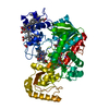
 Basic information
 Components
 Keywords
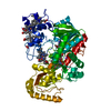
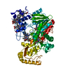
 OXIDOREDUCTASE /
  Function and homology information
 Authors
 Citation
 Structure visualization
 Downloads & links
 Other downloads
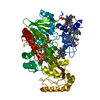


 PDBj
PDBj


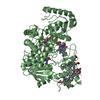
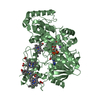
 Assembly
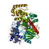


 Mass: 618.503 Da / Num. of mol.: 4 / Source method: obtained synthetically / Formula: C34H34FeN4O4
 Mass: 785.550 Da / Num. of mol.: 1 / Source method: obtained synthetically / Formula: C27H33N9O15P2 / Comment: FAD*YM
 Mass: 132.072 Da / Num. of mol.: 1 / Source method: obtained synthetically / Formula: C4H4O5
 Mass: 22.990 Da / Num. of mol.: 1 / Source method: obtained synthetically / Formula: Na
 Mass: 92.094 Da / Num. of mol.: 1 / Source method: obtained synthetically / Formula: C3H8O3
 Sample preparation
 / Beamline: PX7.2 / Wavelength: 1.488, 1.736, 1.738
 Processing