| 登録情報 | データベース: PDB / ID: 2q12
|
|---|
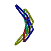
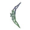
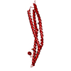
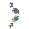
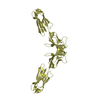
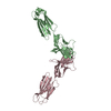
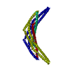
| タイトル | Crystal Structure of BAR domain of APPL1 |
|---|
 要素 要素 | DCC-interacting protein 13 alpha |
|---|
 キーワード キーワード | PROTEIN TRANSPORT / APPL1 / BAR domain |
|---|
| 機能・相同性 |  機能・相同性情報 機能・相同性情報
negative regulation of Fc-gamma receptor signaling pathway involved in phagocytosis / positive regulation of macropinocytosis / adiponectin-activated signaling pathway / macropinosome / regulation of fibroblast migration / regulation of glucose import / protein kinase B binding / maintenance of synapse structure / signaling / positive regulation of melanin biosynthetic process ...negative regulation of Fc-gamma receptor signaling pathway involved in phagocytosis / positive regulation of macropinocytosis / adiponectin-activated signaling pathway / macropinosome / regulation of fibroblast migration / regulation of glucose import / protein kinase B binding / maintenance of synapse structure / signaling / positive regulation of melanin biosynthetic process / intracellular vesicle / regulation of toll-like receptor 4 signaling pathway / early phagosome / positive regulation of cytokine production involved in inflammatory response / Caspase activation via Dependence Receptors in the absence of ligand / vesicle membrane / cellular response to hepatocyte growth factor stimulus / regulation of innate immune response / beta-tubulin binding / phosphatidylserine binding / regulation of G1/S transition of mitotic cell cycle / regulation of protein localization to plasma membrane / ruffle / phosphatidylinositol binding / transforming growth factor beta receptor signaling pathway / positive regulation of glucose import / protein import into nucleus / insulin receptor signaling pathway / early endosome membrane / presynapse / cytoplasmic vesicle / postsynapse / endosome / endosome membrane / early endosome / glutamatergic synapse / protein-containing complex binding / signal transduction / protein homodimerization activity / extracellular exosome / identical protein binding / membrane / nucleus / plasma membrane / cytoplasm / cytosol類似検索 - 分子機能 APPL1, BAR domain / : / : / : / BAR domain of APPL family / Arfaptin homology (AH) domain/BAR domain / BAR domain / Phosphotyrosine interaction domain (PTB/PID) / Phosphotyrosine interaction domain (PID) profile. / Phosphotyrosine-binding domain, phosphotyrosine-interaction (PI) domain ...APPL1, BAR domain / : / : / : / BAR domain of APPL family / Arfaptin homology (AH) domain/BAR domain / BAR domain / Phosphotyrosine interaction domain (PTB/PID) / Phosphotyrosine interaction domain (PID) profile. / Phosphotyrosine-binding domain, phosphotyrosine-interaction (PI) domain / PTB/PI domain / AH/BAR domain superfamily / Substrate Binding Domain Of Dnak; Chain:A; Domain 2 / PH domain / PH domain profile. / Pleckstrin homology domain. / Pleckstrin homology domain / PH-like domain superfamily / Up-down Bundle / Mainly Alpha類似検索 - ドメイン・相同性 |
|---|
| 生物種 |  Homo sapiens (ヒト) Homo sapiens (ヒト) |
|---|
| 手法 |  X線回折 / シンクロトロン / 単波長異常分散 / 解像度: 1.79 Å X線回折 / シンクロトロン / 単波長異常分散 / 解像度: 1.79 Å |
|---|
 データ登録者 データ登録者 | Zhang, X.C. / Zhu, G. |
|---|
 引用 引用 | ジャーナル: Embo J. / 年: 2007
タイトル: Structure of the APPL1 BAR-PH domain and characterization of its interaction with Rab5.
著者: Zhu, G. / Chen, J. / Liu, J. / Brunzelle, J.S. / Huang, B. / Wakeham, N. / Terzyan, S. / Li, X. / Rao, Z. / Li, G. / Zhang, X.C. |
|---|
| 履歴 | | 登録 | 2007年5月23日 | 登録サイト: RCSB / 処理サイト: RCSB |
|---|
| 改定 1.0 | 2007年8月14日 | Provider: repository / タイプ: Initial release |
|---|
| 改定 1.1 | 2011年7月13日 | Group: Derived calculations / Version format compliance |
|---|
|
|---|
 ムービー
ムービー コントローラー


 データを開く
 基本情報
基本情報 構造の表示
構造の表示 ダウンロードとリンク
 その他のダウンロード

 PDBj
PDBj


 集合体
集合体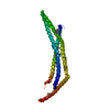

 分子量: 18.015 Da / 分子数: 147 / 由来タイプ: 天然 / 式: H2O
 試料調製
試料調製 / ビームライン: 22-BM / 波長: 0.9793 Å
 解析
解析