 ムービー
ムービー コントローラー


+ データを開く

- 基本情報
基本情報
| 登録情報 | データベース: EMDB / ID: EMD-6301 | |||||||||
|---|---|---|---|---|---|---|---|---|---|---|
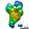
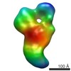
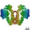
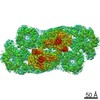
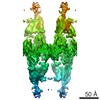
| タイトル | S. cerevisiae SAGA complex in the donut conformation | |||||||||
 マップデータ マップデータ | 3D reconstruction of the donut conformation of SAGA | |||||||||
 試料 試料 |
| |||||||||
 キーワード キーワード |  Histone acetyltransferase (ヒストンアセチルトランスフェラーゼ) Histone acetyltransferase (ヒストンアセチルトランスフェラーゼ) | |||||||||
| 生物種 |  Saccharomyces cerevisiae (パン酵母) Saccharomyces cerevisiae (パン酵母) | |||||||||
| 手法 | 単粒子再構成法 / ネガティブ染色法 / 解像度: 38.9 Å | |||||||||
 データ登録者 データ登録者 | Setiaputra D / Ross JD / Lu S / Cheng DT / Dong MQ / Yip CK | |||||||||
 引用 引用 | ジャーナル: J Biol Chem / 年: 2015 タイトル: Conformational flexibility and subunit arrangement of the modular yeast Spt-Ada-Gcn5 acetyltransferase complex. 著者: Dheva Setiaputra / James D Ross / Shan Lu / Derrick T Cheng / Meng-Qiu Dong / Calvin K Yip /   要旨: The Spt-Ada-Gcn5 acetyltransferase (SAGA) complex is a highly conserved, 19-subunit histone acetyltransferase complex that activates transcription through acetylation and deubiquitination of ...The Spt-Ada-Gcn5 acetyltransferase (SAGA) complex is a highly conserved, 19-subunit histone acetyltransferase complex that activates transcription through acetylation and deubiquitination of nucleosomal histones in Saccharomyces cerevisiae. Because SAGA has been shown to display conformational variability, we applied gradient fixation to stabilize purified SAGA and systematically analyzed this flexibility using single-particle EM. Our two- and three-dimensional studies show that SAGA adopts three major conformations, and mutations of specific subunits affect the distribution among these. We also located the four functional modules of SAGA using electron microscopy-based labeling and transcriptional activator binding analyses and show that the acetyltransferase module is localized in the most mobile region of the complex. We further comprehensively mapped the subunit interconnectivity of SAGA using cross-linking mass spectrometry, revealing that the Spt and Taf subunits form the structural core of the complex. These results provide the necessary restraints for us to generate a model of the spatial arrangement of all SAGA subunits. According to this model, the chromatin-binding domains of SAGA are all clustered in one face of the complex that is highly flexible. Our results relate information of overall SAGA structure with detailed subunit level interactions, improving our understanding of its architecture and flexibility. | |||||||||
| 履歴 |
|
- 構造の表示
構造の表示
| ムービー |
 ムービービューア ムービービューア |
|---|---|
| 構造ビューア | EMマップ: SurfViewMolmilJmol/JSmol |
| 添付画像 |
- ダウンロードとリンク
-EMDBアーカイブ
| マップデータ | emd_6301.map.gz | 7.4 MB | EMDBマップデータ形式 | |
|---|---|---|---|---|
| ヘッダ (付随情報) | emd-6301-v30.xmlemd-6301.xml | 9.8 KB 9.8 KB | 表示 表示 | EMDBヘッダ |
| 画像 |  400_6301.gif 400_6301.gif 80_6301.gif 80_6301.gif | 48.9 KB 3.7 KB | ||
| アーカイブディレクトリ |  http://ftp.pdbj.org/pub/emdb/structures/EMD-6301ftp://ftp.pdbj.org/pub/emdb/structures/EMD-6301 http://ftp.pdbj.org/pub/emdb/structures/EMD-6301ftp://ftp.pdbj.org/pub/emdb/structures/EMD-6301 | HTTPS FTP |
-関連構造データ
-リンク
| EMDBのページ | EMDB (EBI/PDBe) / EMDataResource |
|---|
-マップ
| ファイル | ダウンロード / ファイル: emd_6301.map.gz / 形式: CCP4 / 大きさ: 7.8 MB / タイプ: IMAGE STORED AS FLOATING POINT NUMBER (4 BYTES) | ||||||||||||||||||||||||||||||||||||||||||||||||||||||||||||
|---|---|---|---|---|---|---|---|---|---|---|---|---|---|---|---|---|---|---|---|---|---|---|---|---|---|---|---|---|---|---|---|---|---|---|---|---|---|---|---|---|---|---|---|---|---|---|---|---|---|---|---|---|---|---|---|---|---|---|---|---|---|
| 注釈 | 3D reconstruction of the donut conformation of SAGA | ||||||||||||||||||||||||||||||||||||||||||||||||||||||||||||
| ボクセルのサイズ | X=Y=Z: 4.67 Å | ||||||||||||||||||||||||||||||||||||||||||||||||||||||||||||
| 密度 |
| ||||||||||||||||||||||||||||||||||||||||||||||||||||||||||||
| 対称性 | 空間群: 1 | ||||||||||||||||||||||||||||||||||||||||||||||||||||||||||||
| 詳細 | EMDB XML:
CCP4マップ ヘッダ情報:
| ||||||||||||||||||||||||||||||||||||||||||||||||||||||||||||
-添付データ
- 試料の構成要素
-全体 : The donut conformation of the yeast SAGA complex
| 全体 | 名称: The donut conformation of the yeast SAGA complex |
|---|---|
| 要素 |
|
-超分子 #1000: The donut conformation of the yeast SAGA complex
| 超分子 | 名称: The donut conformation of the yeast SAGA complex / タイプ: sample / ID: 1000 / Number unique components: 1 |
|---|---|
| 分子量 | 理論値: 1.7 MDa |
-分子 #1: Spt-Ada-Gcn5 Acetyltransferase Complex
| 分子 | 名称: Spt-Ada-Gcn5 Acetyltransferase Complex / タイプ: protein_or_peptide / ID: 1 / Name.synonym: SAGA 詳細: Samples were prepared using gradient fixation using a glycerol gradient and glutaraldehyde. 組換発現: No / データベース: NCBI |
|---|---|
| 由来(天然) | 生物種: Saccharomyces cerevisiae (パン酵母) / 株: SF1 / 別称: Yeast / 細胞中の位置: Nucleus |
| 分子量 | 理論値: 1.7 MDa |
-実験情報
-構造解析
| 手法 | ネガティブ染色法 |
|---|---|
 解析 解析 | 単粒子再構成法 |
| 試料の集合状態 | particle |
-試料調製
| 緩衝液 | pH: 7.4 詳細: 40 mM HEPES, 150 mM NaCl, 20% glycerol, 0.1% Tween-20, 0.015% glutaraldehyde |
|---|---|
| 染色 | タイプ: NEGATIVE 詳細: Grids with adsorbed protein were used to scoop a carbon layer floating on 0.75% w/v uranyl formate, sandwiching the protein and a layer of stain between two carbon layers. |
| グリッド | 詳細: 400 mesh copper grid with holey carbon support, glow discharged |
| 凍結 | 凍結剤: NONE / 装置: OTHER |
- 電子顕微鏡法
| 顕微鏡 | FEI TECNAI SPIRIT |
|---|---|
| 電子線 | 加速電圧: 120 kV / 電子線源: LAB6 |
| 電子光学系 | 倍率(補正後): 65900 / 照射モード: FLOOD BEAM / 撮影モード: BRIGHT FIELDBright-field microscopy / 最大 デフォーカス(公称値): 1.2 µm / 最小 デフォーカス(公称値): 0.8 µm / 倍率(公称値): 49000 |
| 試料ステージ | 試料ホルダー: Side entry room temperature tomography holder 試料ホルダーモデル: SIDE ENTRY, EUCENTRIC / Tilt angle min: -65 |
| 温度 | 平均: 298 K |
| 詳細 | Low dose |
| 日付 | 2013年2月10日 |
| 撮影 | カテゴリ: CCD / フィルム・検出器のモデル: FEI EAGLE (4k x 4k) / デジタル化 - サンプリング間隔: 1.3 µm / 実像数: 382 / 平均電子線量: 25 e/Å2 / ビット/ピクセル: 16 |
| Tilt angle max | 0 |
| 実験機器 |  モデル: Tecnai Spirit / 画像提供: FEI Company |
-画像解析
| 最終 再構成 | アルゴリズム: OTHER / 解像度のタイプ: BY AUTHOR / 解像度: 38.9 Å / 解像度の算出法: OTHER / ソフトウェア - 名称: Spider 詳細: The initial map was made using back-projection of tilted data, and was subjected to an angular refinement cycle with the same data set. The model then underwent a final refinement cycle with ...詳細: The initial map was made using back-projection of tilted data, and was subjected to an angular refinement cycle with the same data set. The model then underwent a final refinement cycle with both tilted and untilted data. The model was then filtered to the calculated resolution. 使用した粒子像数: 22518 |
|---|---|
| 詳細 | Particles were selected manually. |
