National Institutes of Health/National Cancer Institute
R01CA193578
米国
National Institutes of Health/National Institute Of Allergy and Infectious Diseases
R01AI116815
米国
引用
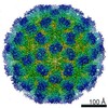
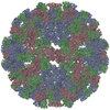
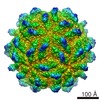
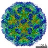
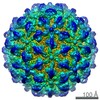
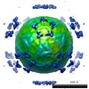
ジャーナル: Comput Struct Biotechnol J / 年: 2019 タイトル: Cryo-EM Reveals Architectural Diversity in Active Rotavirus Particles. 著者: Mary Hauser / William J Dearnaley / A Cameron Varano / Michael Casasanta / Sarah M McDonald / Deborah F Kelly / 要旨: Rotavirus is a well-studied RNA virus that causes severe gastroenteritis in children. During viral entry, the outer layer of the virion is shed, creating a double-layered particle (DLP) that is ...Rotavirus is a well-studied RNA virus that causes severe gastroenteritis in children. During viral entry, the outer layer of the virion is shed, creating a double-layered particle (DLP) that is competent to perform viral transcription (i.e., mRNA synthesis) and launch infection. While inactive forms of rotavirus DLPs have been structurally characterized in detail, information about the transcriptionally-active DLP remains limited. Here, we used cryo-Electron Microscopy (cryo-EM) and 3D image reconstructions to compare the structures of internal protein components in transcriptionally-active versus inactive DLPs. Our findings showed that transcriptionally-active DLPs gained internal order as mRNA synthesis unfolded, while inactive DLPs remained dynamically disordered. Regions of viral protein/RNA constituents were analyzed across two different axes of symmetry to provide a more comprehensive view of moving components. Taken together, our results bring forth a new view of active DLPs, which may enable future pharmacological strategies aimed at obliterating rotavirus transcription as a therapeutic approach.
 ムービー
ムービー コントローラー


 データを開く
 基本情報
基本情報 マップデータ
 試料
試料 Simian rotavirus A/SA11-4F (ウイルス)
 データ登録者
 米国, 2件
  引用
引用 構造の表示
構造の表示 ムービービューア
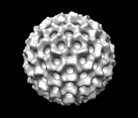
 ダウンロードとリンク
 emd_20592.png
 http://ftp.pdbj.org/pub/emdb/structures/EMD-20592
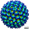
 Z (Sec.)
 Y (Row.)
 X (Col.)


 試料の構成要素
 解析
解析 電子顕微鏡法
 FIELD EMISSION GUN
