ジャーナル: J Virol / 年: 2011 タイトル: The herpes simplex virus 1 UL17 protein is the second constituent of the capsid vertex-specific component required for DNA packaging and retention. 著者: Katerina Toropova / Jamie B Huffman / Fred L Homa / James F Conway / 要旨: The herpes simplex virus (HSV) UL17 and UL25 minor capsid proteins are essential for DNA packaging. They are thought to comprise a molecule arrayed in five copies around each of the capsid vertices. ...The herpes simplex virus (HSV) UL17 and UL25 minor capsid proteins are essential for DNA packaging. They are thought to comprise a molecule arrayed in five copies around each of the capsid vertices. This molecule was initially termed the "C-capsid-specific component" (CCSC) (B. L. Trus et al., Mol. Cell 26:479-489, 2007), but as we have subsequently observed this feature on reconstructions of A, B, and C capsids, we now refer to it more generally as the "capsid vertex-specific component" (CVSC) (S. K. Cockrell et al., J. Virol. 85:4875-4887, 2011). We previously confirmed that UL25 occupies the vertex-distal region of the CVSC density by visualizing a large UL25-specific tag in reconstructions calculated from cryo-electron microscopy (cryo-EM) images. We have pursued the same strategy to determine the capsid location of the UL17 protein. Recombinant viruses were generated that contained either a small tandem affinity purification (TAP) tag or the green fluorescent protein (GFP) attached to the C terminus of UL17. Purification of the TAP-tagged UL17 or a similarly TAP-tagged UL25 protein clearly demonstrated that the two proteins interact. A cryo-EM reconstruction of capsids containing the UL17-GFP protein reveals that UL17 is the second component of the CVSC and suggests that UL17 interfaces with the other CVSC component, UL25, through its C terminus. The portion of UL17 nearest the vertex appears to be poorly constrained, which may provide flexibility in interacting with tegument proteins or the DNA-packaging machinery at the portal vertex. The exposed locations of the UL17 and UL25 proteins on the HSV-1 capsid exterior suggest that they may be attractive targets for highly specific antivirals.
ダウンロード / ファイル: emd_1903.map.gz / 形式: CCP4 / 大きさ: 985.7 MB / タイプ: IMAGE STORED AS FLOATING POINT NUMBER (4 BYTES)
注釈
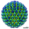
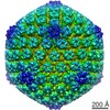
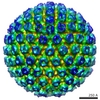
Surface rendered view of a HSV-1 C-capsid where protein UL17 has been genetically tagged with GFP at its C-terminus
ボクセルのサイズ
X=Y=Z: 2.12 Å
密度
表面レベル
登録者による: 0.7 / ムービー #1: 0.7
最小 - 最大
-6.45088243 - 5.97226095
平均 (標準偏差)
-0.00000002 (±1.0)
対称性
空間群: 1
詳細
EMDB XML:
マップ形状
Axis order
X
Y
Z
Origin
0
0
0
サイズ
642
642
642
Spacing
642
642
642
セル
A=B=C: 1361.0399 Å α=β=γ: 90.0 °
CCP4マップ ヘッダ情報:
mode
Image stored as Reals
Å/pix. X/Y/Z
2.12
2.12
2.12
M x/y/z
642
642
642
origin x/y/z
0.000
0.000
0.000
length x/y/z
1361.040
1361.040
1361.040
α/β/γ
90.000
90.000
90.000
start NX/NY/NZ
-56
-56
-55
NX/NY/NZ
112
112
112
MAP C/R/S
1
2
3
start NC/NR/NS
0
0
0
NC/NR/NS
642
642
642
D min/max/mean
-6.451
5.972
-0.000
-
添付データ
-
試料の構成要素
-
全体 : HSV-1 C-capsid with UL17 protein labeled with GFP at its C terminus.
全体
名称: HSV-1 C-capsid with UL17 protein labeled with GFP at its C terminus.
要素
試料: HSV-1 C-capsid with UL17 protein labeled with GFP at its C terminus.
ウイルス: Human herpesvirus 1 strain KOS (ヘルペスウイルス)
-
超分子 #1000: HSV-1 C-capsid with UL17 protein labeled with GFP at its C terminus.
超分子
名称: HSV-1 C-capsid with UL17 protein labeled with GFP at its C terminus. タイプ: sample / ID: 1000 / Number unique components: 1
-
超分子 #1: Human herpesvirus 1 strain KOS
超分子
名称: Human herpesvirus 1 strain KOS / タイプ: virus / ID: 1 / Name.synonym: HSV-1 / NCBI-ID: 10306 / 生物種: Human herpesvirus 1 strain KOS / ウイルスタイプ: VIRION / ウイルス・単離状態: STRAIN / ウイルス・エンベロープ: No / ウイルス・中空状態: No / Syn species name: HSV-1
宿主
生物種: Homo sapiens (ヒト) / 別称: VERTEBRATES
ウイルス殻
Shell ID: 1 / 直径: 1250 Å / T番号(三角分割数): 16
-
実験情報
-
構造解析
手法
クライオ電子顕微鏡法
解析
単粒子再構成法
試料の集合状態
particle
-
試料調製
緩衝液
pH: 7.5 / 詳細: 500 mM NaCl, 10 mM Tris, 1 mM EDTA
凍結
凍結剤: OTHER / チャンバー内湿度: 85 % / 装置: FEI VITROBOT MARK III 詳細: Vitrification instrument: Vitrobot mark III. Cryogen was an equal mix of ethane - propane 手法: 7 second blot before plunging
 ムービー
ムービー コントローラー


 データを開く
 基本情報
基本情報 マップデータ
 試料
試料 キーワード
キーワード HSV-1 (単純ヘルペスウイルス) / UL17 / UL25 / CVSC /
 データ登録者
 引用
引用
 構造の表示
構造の表示 ムービービューア
 ダウンロードとリンク
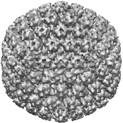 EMD-1903.png
 http://ftp.pdbj.org/pub/emdb/structures/EMD-1903
 試料の構成要素
 解析
解析 電子顕微鏡法
