 ムービー
ムービー コントローラー


+ データを開く

- 基本情報
基本情報
| 登録情報 |  | |||||||||
|---|---|---|---|---|---|---|---|---|---|---|
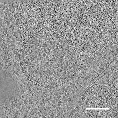
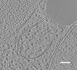
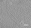
| タイトル | In situ cryo-electron tomogram of a bulk autophagy autophagosome fusing with the vacuole in S. cerevisiae #1 | |||||||||
 マップデータ マップデータ | Fusion of autophagosome with the vacuole, in situ tomogram in S. cerevisiae | |||||||||
 試料 試料 |
| |||||||||
 キーワード キーワード |  Autophagy (オートファジー) / phagophore / fusion / yeast (酵母) / bulk / degradation / RIBOSOME (リボソーム) Autophagy (オートファジー) / phagophore / fusion / yeast (酵母) / bulk / degradation / RIBOSOME (リボソーム) | |||||||||
| 生物種 |  Saccharomyces cerevisiae (パン酵母) Saccharomyces cerevisiae (パン酵母) | |||||||||
| 手法 | 電子線トモグラフィー法 / クライオ電子顕微鏡法 | |||||||||
 データ登録者 データ登録者 | Bieber A / Capitanio C / Erdmann PS / Schulman BA / Baumeister W / Wilfling F | |||||||||
| 資金援助 |  米国, 1件 米国, 1件
| |||||||||
 引用 引用 | ジャーナル: Proc Natl Acad Sci U S A / 年: 2022 タイトル: In situ structural analysis reveals membrane shape transitions during autophagosome formation. 著者: Anna Bieber / Cristina Capitanio / Philipp S Erdmann / Fabian Fiedler / Florian Beck / Chia-Wei Lee / Delong Li / Gerhard Hummer / Brenda A Schulman / Wolfgang Baumeister / Florian Wilfling /   要旨: Autophagosomes are unique organelles that form de novo as double-membrane vesicles engulfing cytosolic material for destruction. Their biogenesis involves membrane transformations of distinctly ...Autophagosomes are unique organelles that form de novo as double-membrane vesicles engulfing cytosolic material for destruction. Their biogenesis involves membrane transformations of distinctly shaped intermediates whose ultrastructure is poorly understood. Here, we combine cell biology, correlative cryo-electron tomography (cryo-ET), and extensive data analysis to reveal the step-by-step structural progression of autophagosome biogenesis at high resolution directly within yeast cells. The analysis uncovers an unexpectedly thin intermembrane distance that is dilated at the phagophore rim. Mapping of individual autophagic structures onto a timeline based on geometric features reveals a dynamical change of membrane shape and curvature in growing phagophores. Moreover, our tomograms show the organelle interactome of growing autophagosomes, highlighting a polar organization of contact sites between the phagophore and organelles, such as the vacuole and the endoplasmic reticulum (ER). Collectively, these findings have important implications for the contribution of different membrane sources during autophagy and for the forces shaping and driving phagophores toward closure without a templating cargo. | |||||||||
| 履歴 |
|
- 構造の表示
構造の表示
| 添付画像 |
|---|
- ダウンロードとリンク
-EMDBアーカイブ
| マップデータ | emd_15547.map.gz | 761.3 MB |  EMDBマップデータ形式 EMDBマップデータ形式 | |
|---|---|---|---|---|
| ヘッダ (付随情報) | emd-15547-v30.xmlemd-15547.xml | 11.4 KB 11.4 KB | 表示 表示 | EMDBヘッダ |
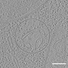
| 画像 |  emd_15547.png emd_15547.png | 265.3 KB | ||
| Filedesc metadata | emd-15547.cif.gz | 4.1 KB | ||
| アーカイブディレクトリ |  http://ftp.pdbj.org/pub/emdb/structures/EMD-15547ftp://ftp.pdbj.org/pub/emdb/structures/EMD-15547 http://ftp.pdbj.org/pub/emdb/structures/EMD-15547ftp://ftp.pdbj.org/pub/emdb/structures/EMD-15547 | HTTPS FTP |
-関連構造データ
-リンク
| EMDBのページ | EMDB (EBI/PDBe) / EMDataResource |
|---|
-マップ
| ファイル | ダウンロード / ファイル: emd_15547.map.gz / 形式: CCP4 / 大きさ: 821.3 MB / タイプ: IMAGE STORED AS FLOATING POINT NUMBER (4 BYTES) | ||||||||||||||||||||
|---|---|---|---|---|---|---|---|---|---|---|---|---|---|---|---|---|---|---|---|---|---|
| 注釈 | Fusion of autophagosome with the vacuole, in situ tomogram in S. cerevisiae | ||||||||||||||||||||
| ボクセルのサイズ | X=Y=Z: 14.08 Å | ||||||||||||||||||||
| 密度 |
| ||||||||||||||||||||
| 対称性 | 空間群: 1 | ||||||||||||||||||||
| 詳細 | EMDB XML:
|
-添付データ
- 試料の構成要素
-全体 : In situ cryo-electron tomogram of a bulk autophagy autophagosome ...
| 全体 | 名称: In situ cryo-electron tomogram of a bulk autophagy autophagosome fusing with the vacuole in S. cerevisiae #1 |
|---|---|
| 要素 |
|
-超分子 #1: In situ cryo-electron tomogram of a bulk autophagy autophagosome ...
| 超分子 | 名称: In situ cryo-electron tomogram of a bulk autophagy autophagosome fusing with the vacuole in S. cerevisiae #1 タイプ: cell / ID: 1 / 親要素: 0 詳細: Autophagosome fusing with the vacuole. The position of the autophagosome corresponds to mCherry-Atg8 signal detected by cryo-fluorescence microscopy. |
|---|---|
| 由来(天然) | 生物種: Saccharomyces cerevisiae (パン酵母) / 株: DF5 |
-実験情報
-構造解析
| 手法 | クライオ電子顕微鏡法 |
|---|---|
 解析 解析 | 電子線トモグラフィー法 |
| 試料の集合状態 | cell |
-試料調製
| 緩衝液 | pH: 7 |
|---|---|
| グリッド | モデル: Quantifoil / 支持フィルム - トポロジー: HOLEY |
| 凍結 | 凍結剤: ETHANE-PROPANE / チャンバー内温度: 295 K / 装置: FEI VITROBOT MARK IV |
| 詳細 | S. cerevisiae. 2 hours starvation in SD-N. |
| 切片作成 | 集束イオンビーム - 装置: OTHER / 集束イオンビーム - イオン: OTHER / 集束イオンビーム - 電圧: 30 / 集束イオンビーム - 電流: 0.3 / 集束イオンビーム - 時間: 2400 / 集束イオンビーム - 温度: 91 K / 集束イオンビーム - Initial thickness: 7000 / 集束イオンビーム - 最終 厚さ: 150 集束イオンビーム - 詳細: Correlative FIB-milling. The lamella was prepared in the location of fluorescence signal.. The value given for _em_focused_ion_beam.instrument is Other. This is not ...集束イオンビーム - 詳細: Correlative FIB-milling. The lamella was prepared in the location of fluorescence signal.. The value given for _em_focused_ion_beam.instrument is Other. This is not in a list of allowed values {'DB235', 'OTHER'} so OTHER is written into the XML file. |
- 電子顕微鏡法
| 顕微鏡 | FEI TITAN KRIOS |
|---|---|
| 電子線 | 加速電圧: 300 kV / 電子線源: FIELD EMISSION GUN |
| 電子光学系 | 照射モード: FLOOD BEAM / 撮影モード: BRIGHT FIELDBright-field microscopy / 最大 デフォーカス(公称値): 4.5 µm / 最小 デフォーカス(公称値): 4.0 µm / 倍率(公称値): 42000 |
| 試料ステージ | 試料ホルダーモデル: FEI TITAN KRIOS AUTOGRID HOLDER ホルダー冷却材: NITROGEN |
| 撮影 | フィルム・検出器のモデル: GATAN K2 SUMMIT (4k x 4k) 検出モード: COUNTING / デジタル化 - サイズ - 横: 3712 pixel / デジタル化 - サイズ - 縦: 3712 pixel / 実像数: 60 / 平均露光時間: 2.0 sec. / 平均電子線量: 120.0 e/Å2 |
| 実験機器 |  モデル: Titan Krios / 画像提供: FEI Company |
-画像解析
| 最終 再構成 | ソフトウェア - 名称: IMOD / 使用した粒子像数: 55 |
|---|
