ジャーナル: EMBO J / 年: 2022 タイトル: Cooperative amyloid fibre binding and disassembly by the Hsp70 disaggregase. 著者: Joseph George Beton / Jim Monistrol / Anne Wentink / Erin C Johnston / Anthony John Roberts / Bernd Gerhard Bukau / Bart W Hoogenboom / Helen R Saibil / 要旨: Although amyloid fibres are highly stable protein aggregates, a specific combination of human Hsp70 system chaperones can disassemble them, including fibres formed of α-synuclein, huntingtin, or Tau. ...Although amyloid fibres are highly stable protein aggregates, a specific combination of human Hsp70 system chaperones can disassemble them, including fibres formed of α-synuclein, huntingtin, or Tau. Disaggregation requires the ATPase activity of the constitutively expressed Hsp70 family member, Hsc70, together with the J domain protein DNAJB1 and the nucleotide exchange factor Apg2. Clustering of Hsc70 on the fibrils appears to be necessary for disassembly. Here we use atomic force microscopy to show that segments of in vitro assembled α-synuclein fibrils are first coated with chaperones and then undergo bursts of rapid, unidirectional disassembly. Cryo-electron tomography and total internal reflection fluorescence microscopy reveal fibrils with regions of densely bound chaperones, preferentially at one end of the fibre. Sub-stoichiometric amounts of Apg2 relative to Hsc70 dramatically increase recruitment of Hsc70 to the fibres, creating localised active zones that then undergo rapid disassembly at a rate of ~ 4 subunits per second. The observed unidirectional bursts of Hsc70 loading and unravelling may be explained by differences between the two ends of the polar fibre structure.
 ムービー
ムービー コントローラー


 データを開く
 基本情報
基本情報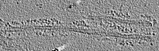
 マップデータ
 試料
試料
 Homo sapiens (ヒト)
 データ登録者
 英国, 2件
  引用
引用
 構造の表示
構造の表示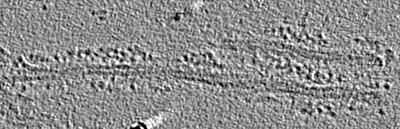
 ダウンロードとリンク
 EMDBマップデータ形式
 emd_13576.png
 http://ftp.pdbj.org/pub/emdb/structures/EMD-13576
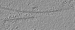
 試料の構成要素
 解析
解析 電子顕微鏡法
