凍結剤: ETHANE-PROPANE / 装置: HOMEMADE PLUNGER 詳細: The grids were mounted on a manual plunger, blotted from the back side using Whatman paper #1 (Sigma-Aldrich) and plunged into a 2:1 ethane:propane mixture cooled down by liquid nitrogen by ...詳細: The grids were mounted on a manual plunger, blotted from the back side using Whatman paper #1 (Sigma-Aldrich) and plunged into a 2:1 ethane:propane mixture cooled down by liquid nitrogen by the Martinsried-Plunger..
詳細
Dissected intact Drosophila body wall muscles were deposited on an EM grid.
Cryo protectant
10% glycerol
切片作成
集束イオンビーム - 装置: OTHER / 集束イオンビーム - イオン: OTHER / 集束イオンビーム - 電圧: 30 kV / 集束イオンビーム - 電流: 0.05 nA / 集束イオンビーム - 時間: 3600 sec. / 集束イオンビーム - 温度: 90 K / 集束イオンビーム - Initial thickness: 60000 nm / 集束イオンビーム - 最終 厚さ: 180 nm 集束イオンビーム - 詳細: To prepare thin electron transparent lamellae into the tissue, plunge-frozen grids were first mounted into Autogrid frames (FEI). The grids were then mounted into a ...集束イオンビーム - 詳細: To prepare thin electron transparent lamellae into the tissue, plunge-frozen grids were first mounted into Autogrid frames (FEI). The grids were then mounted into a dual-beam Quanta 3D FIB/SEM (FEI) using a custom-built transfer shuttle and a cryo-transfer system (PP3000T, Quorum). The samples were kept at -180 C throughout FIB milling by the cryo-stage. To improve SEM imaging, a thin layer of pure metallic Pt was sputtered onto the sample under cryo conditions in the PP3000T transfer system to increase its electrical conductivity. The following parameters were used: 10 mA sputtering current, 500 V between stage and sputtering target and 30 s of exposure at 4.5x10⁻² mbar. To interpret and annotate the topographical anatomy of the tissue, overview maps of the EM grid were acquired by SEM at 10 kV at 100-250x magnification (object pixel size 1.1-0.4 um) and by secondary electrons induced by the Ga+ focused ion beam at 30 kV at 338x magnification (object pixel size 0.7 um). To protect the milling front of the lamellae, gaseous organic platinum was frozen on top of the grid using a gas injection system. To prevent bending of the lamella during the preparation, micro-expansion joints were milled left and right of the intended lamella preparation site. 10-20 um wide lamellae were prepared into the tissue with the ion beam at 30 kV at shallow angles (8-14 deg) in four consecutive steps: for the thicker tissue regions (e.g. VNC or skeletal muscle), the areas above and below the intended lamella were first removed with an ion beam current of 5 nA and 10 um spacing. This step was not necessary for thinner tissues such as the peripheral nerves. Further rectangular patterns were defined above and below the intended lamella with 2 um spacing for the rough milling step (ion beam current of 500-1000 pA), followed by fine milling with 800 nm spacing (100 pA) and a final polishing step down to the final lamella thickness of 100-200 nm (50 pA). To reach a uniform thickness, the lamella was tilted by +-0.5 deg and milled on each side separately with 50 pA current. The thickness of the lamella during the polishing step was assessed by SEM at 3-5 kV, 4.1 pA: the loss of charging effects in the lamella, visualized as the vanishing of bright areas, indicates a thickness <300 nm at 5 kV or <200 nm at 3 kV. Biological structures inside the lamella at the surface were imaged at each step by SEM at 2.5 kV, 4.1 pA in integration mode (64x). To reduce lamella charging during phase plate cryo-ET data acquisition, a thin layer of pure metallic Pt was sputtered onto the lamella under cryo conditions in the PP3000T transfer system with the following parameters: 5 mA sputtering current, 500 V between stage and sputtering target and 10 s of exposure at 4.5x10⁻² mbar.. The value given for _emd_sectioning_focused_ion_beam.instrument is FEI Quanta 3D FIB/SEM. This is not in a list of allowed values {'DB235', 'OTHER'} so OTHER is written into the XML file.
 ムービー
ムービー コントローラー


 データを開く
 基本情報
基本情報 マップデータ
 試料
試料
 Drosophila melanogaster (キイロショウジョウバエ)
 データ登録者
 ドイツ,
ドイツ,  中国, 3件
  引用
引用 構造の表示
構造の表示 ムービービューア
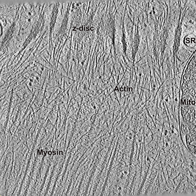
 ダウンロードとリンク
 emd_12728.png
 http://ftp.pdbj.org/pub/emdb/structures/EMD-12728
 試料の構成要素
 解析
解析 電子顕微鏡法
